SciELO - Scientific Electronic Library Online

 
vol.52 issue2How to protect Peruvian coastal wetlands? An answer based on a conceptual model of their drivers of changeMass mortality event of the Caribbean sharpnose puffer Canthigaster rostrata (Tetraodontiformes: Tetraodontidae) in the Costa Rican southern Caribbean during 2020 author indexsubject indexarticles search
Home Pagealphabetic serial listing  

Services on Demand

Journal

Article

Indicators

Related links

  • On index processCited by Google
  • Have no similar articlesSimilars in SciELO
  • On index processSimilars in Google

Share


Boletín de Investigaciones Marinas y Costeras - INVEMAR

Print version ISSN 0122-9761

Bol. Invest. Mar. Cost. vol.52 no.2 Santa Marta July/Dec. 2023  Epub July 02, 2024

https://doi.org/10.25268/bimc.invemar.2023.52.2.1231 

Research Articles

Fungal endophytes isolated from mangroves in San Pedro Nature Reserve, Buenaventura

Hongos endófitos aislados de manglares en la Reserva Natural San Pedro, Buenaventura

1Programa de Biología, Universidad del Quindío, Carrera 15 # 12N, Armenia-Quindío, Colombia. osorio.romero17@gmail.com

2Programa de Biología, Universidad del Quindío, Carrera 15 # 12N, Armenia-Quindío, Colombia. yriosa@uqvirtual.edu.co

3Natural Resources Institute Finland (Luke), Latokartanonkaari 9, 00790 Helsinki, Finland. riikka.linnakoski@luke.fi


ABSTRACT

Mangrove trees grow in estuarine zones in tropical and subtropical regions, where they provide important economic, ecological and cultural services. Studies have shown that these trees are important reservoirs of fungal microorganisms, which encompasses a range of morphologically diverse fungi that are either pathogens, endophytes or saprobes, playing a key role in nutrient cycling, host protection against adverse conditions and in many occasions host decline. To determine the diversity of endophytic fungi associated with mangrove trees in the San Pedro Natural Reserve, Buenaventura, Colombia, healthy branches (length approx. 10 cm) of Laguncularia racemosa, Mora oleifera, Pelliciera rhizophorae and Rhizophora racemosa were collected for the respective fungal isolations. DNA was extracted and the Internal Transcribed Spacer (ITS) region was amplified, followed by phylogenetic analyses such as Bayesian (BI), Maximum Likelihood (ML) and Maximum Parsimony (MP). A total of nine genera, Bipolaris, Ciboria, Coprinellus, Cylindrobasidium, Epicoccum, Fusarium, Lasiodiplodia, Neofusicoccum and Neurospora belonging to eight families were identified. Of these, Fusarium, Lasiodiplodia and Neofusicoccum are well known for their high potential to threaten the health of their hosts when conditions are adverse. Endophytic fungi also play an important role in the production of biocompounds for the protection of their hosts.

Keywords: Coastal ecosystems; fungal pathogens; fungus-host association; molecular analysis

RESUMEN

Los árboles de mangle crecen en zonas estuarinas de las regiones tropicales y subtropicales, donde prestan importantes servicios económicos, ecológicos y culturales. Los estudios han demostrado que estos árboles son importantes reservorios de microorganismos fúngicos, que comprenden una serie de hongos morfológicamente diversos incluyendo patógenos, endófitos o saprobios, desempeñando un papel clave en el ciclado de nutrientes, protección del hospedero frente a condiciones adversas y, en muchas ocasiones, el declive de los hospederos. Con el fin de determinar la diversidad de hongos endófitos asociados a árboles de mangle en la Reserva Natural San Pedro, Buenaventura, Colombia; se colectaron ramas sanas (longitud aprox. 10 cm) de Laguncularia racemosa, Mora oleifera, Pelliciera rhizophorae y Rhizophora racemosa para los respectivos aislamientos fúngicos. Se extrajo ADN y se amplificó la región del espaciador transcrito interno (ITS), seguido de análisis filogenéticos tales como Bayesiano (BI), Máxima Verosimilitud (ML) y Máxima Parsimonia (MP). En total se identificaron nueve géneros, Bipolaris, Ciboria, Coprinellus, Cylindrobasidium, Epicoccum, Fusarium, Lasiodiplodia, Neofusicoccum y Neurospora pertenecientes a ocho familias. De estos, Fusarium, Lasiodiplodia y Neofusicoccum son bien conocidos por su alto potencial para amenazar la salud de sus huéspedes cuando sus condiciones son adversas, igualmente los hongos endófitos también cumplen un papel importante en la producción de biocompuestos para la protección de sus hospedaros.

Palabras clave: Análisis molecular; asociación hongo-huésped; ecosistemas costeros; patógenos fúngicos

INTRODUCTION

Mangrove forests are highly productive ecosystems of intertidal areas in tropical and subtropical regions (Spalding et al., 2010; Tomlinson, 2016). Colombia is the third-largest mangrove country in the Latin America, with an approximate area of 371,081 ha. of which 282,835 ha. are on the Pacific coast (Giri et al., 2011).

In the Colombian Pacific, mangrove ecosystems consist mainly of eight species including Avicennia germinans (L.), Conocarpus erectus L., Laguncularia racemosa (L.) C.F. Gaertn., Mora oleifera (Triana ex Hemsl.) Ducke, Pelliciera rhizophorae Planch. and Triana, Rhizophora harrisonii Leechm., Rhizophora mangle L., and Rhizophora racemosa G. Mey. (García, 2010; Cornejo et al., 2014; Mejía et al., 2014). In addition, these species also provide important services to the local communities of the region (Palacios and Cantera, 2017). However, despite these benefits, combinations of human activities and natural events have been widely recognized to be associated with the decline of these trees in this part of the country (Prahl, 1989; Álvarez, 2003; López et al., 2016).

In recent years, the productive potential of mangrove ecosystems has been largely considered due to the metabolic activity of fungal communities (Hyde and Lee, 1995; Rineau et al., 2013; Thatoi et al., 2013). This potential has aroused great interest in investigating associations between mangrove trees and potentially beneficial fungi such as endophytes (Petrini, 1991; Wilson, 1995), which colonize the internal tissues of plants by protecting them from adverse factors (Gilbert et al., 2002; Arnold, 2007), or turning as opportunistic pathogens when plants are under different levels of stress (Slippers and Wingfield, 2007).

Numerous species of endophytic fungi have been reported in Colombia, either from native or non-native symptomatic or asymptomatic plants (e.g., Salazar and Garcia, 2005; Ordóez et al., 2012; Gamboa and Otero, 2016). However, studies on these fungal groups associated with mangrove species in the country remain very limited (Bolívar-Anillo et al., 2015). The objective of this study was to evaluate the presence of fungal endophytes associated with mangrove species in the San Pedro Nature Reserve, Buenaventura, Colombia, and to determine their possible role in disease development based on relevant literature.

STUDY AREA

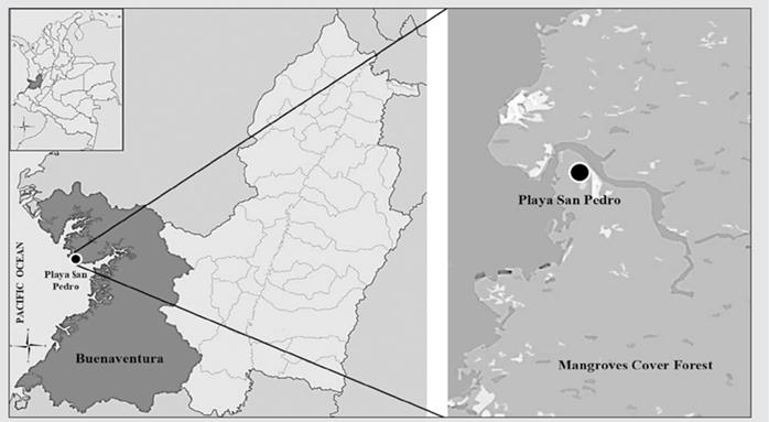
The San Pedro Nature Reserve (03° 50’ 06” N y 77° 15’ 28” W), is located at the isthmus of Pichidó, in the middle area of the Colombian Pacific coast, in the municipality of Buenaventura (Valle del Cauca). The reserve has an area of 100 ha, of which 40 % is mangrove ecosystem and 60 % rainforest. It has an average annual temperature of 26 °C, the relative humidity is 80 % to 98 %, and an average annual rainfall of 7,000 mm. Four mangrove species are present at this nature reserve, including Laguncularia racemosa, Mora oleifera, Pelliciera rhizophorae, and Rhizophora racemosa (Fig. 1).

Figure 1 Study area. San Pedro Nature Reserve, Buenaventura, Valle del Cauca, Colombia. 

MATERIALS AND METHODS

Field sampling

Prior recognition of the study area was performed, followed by a random sampling of asymptomatic branch segments from the middle part of canopy. In total 20 branches of approximately 10 cm long (ca.) were collected, five from each mangrove species including Laguncularia racemosa, Mora oleifera, Pelliciera rhizophorae and Rhizophora racemosa were placed in paper bags and stored in a portable refrigerator. They were subsequently transferred and preserved in the laboratory of the Biology Program of the Universidad del Quindío for further fungal isolation. It needs to be considered that the sampling size (number of branches collected) performed in this study depended on the sampling permits obtain from the National Environmental Licensing Authority.

Fungal isolations

The isolation of endophytes was carried out following the protocol described by Slippers and Wingfield (2007), with certain modifications made by Osorio et al., (2017). The collected branches were washed with tap water to remove excess debris and for each branch, five portions of 0.5 cm approximately were cut. These fragments were submerged in 90 % ethanol for one minute, followed by sodium hypochlorite (NaOCl) at 4 % (1 min), then in 70 % ethanol (1 min), and ending with a rinse in distilled water (1 min).

After surface sterilization and the pieces were placed on sterile paper for drying, each fragment was transferred to 2 % malt extract agar (MEA; 20 g malt extract, 20 g Biolab Agar in 1 L of distilled water), containing 50 mg of streptomycin to prevent bacterial colonization. The samples were incubated at room temperature, in natural light and darkness. The resulting colonies were purified using the “single hyphal tip” method, as described in the procedure carried out by Osorio et al., (2017), subsequently, the resulting colonies were separated into morph-groups for the following DNA extraction, which was performed by following the methodology of Raeder and Broda (1985).

Direct PCR amplification and sequencing

PCR was performed directly from representative pure cultures without prior DNA purification using Phire Plant Direct PCR kit (Thermo Scientific, USA) following the manufacturer’s instructions. ITS1-F and ITS4 primers (White et al., 1990) were used for the amplification of the ITS region. 25 µl PCR reactions contained 0.5 µl DNA template, 0.5 µM ITS1-F and ITS4 primers, 15 µl 2 × Phire Plant PCR buffer (includes dNTPs and MgCl2) (Thermo Scientific) and 0.5 µl Phire Hot Start II DNA Polymerase (Thermo Scientific). Initial denaturation was performed at 98 °C for 5 min followed by 35 cycles of (1) denaturation at 98 °C for 5 s, (2) annealing at 55 °C for 5 s, and (3) extension at 72 °C for 20 s. The final extension occurred at 72 °C for 1 min. The amplified PCR products were run on 1 % agarose gels, and successfully amplified products were purified with EXO-SAP protocol: the PCR product (20 μl) was mixed with 8 μl of Exo-SAP (5 μl of Exonuclease I (20 U/μl) (Fermentas,Vilnius, Lithuania) and 100 μl of Shrimp Alkaline Phosphatase (1 U/μL) (Roche Diagnostics, Indianapolis, USA) in a 1000 μl reaction mixture). The samples were incubated at (1) 37 °C for 15 minutes and (2) at 80 °C for 15 minutes. The PCR products were sequenced at Macrogen Europe. The resulting sequences were manually analyzed, and consensus sequences obtained by using Sequencher v.5 software.

Phylogenetic analysis

The obtained sequences were compared to the species data in GenBank (https://blast.ncbi.nlm.nih.gov/Blast.cgi) by performing BLAST searches on GenBank using the BLASTn algorithm (Altschul et al., 1990). DNA sequences of closely related species were obtained from GenBank and combined into datasets with the ITS sequences of the isolates obtained in this study (Table 1). The selection of the outgroup for each dataset was made according to the results of previous studies, including those of Sun and Guo (2012) and Osorio et al. (2017). The data sets were aligned using the MAFFT program (/), selecting the iterative refinement method G-INS-i (Kuraku et al., 2013; Katoh et al., 2019) and were manually edited with MEGA v. 10.1.7 (Kumar et al., 2018). Phylogenetic reconstructions were performed by maximum parsimony (MP), maximum likelihood (ML), and bayesian inference (BI) for each dataset obtained.

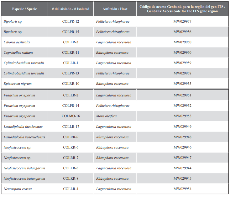
Table 1 Isolate numbers and access information to GenBank of isolates obtained from mangrove trees in the San Pedro Buenaventura Nature Reserve (Colombia). 
Analyses of the MP were performed in PAUP v. 4. 0b10 (Swofford, 2003) using a heuristic search and tree-bisection-reconnection (TBR) reorganization algorithm. Gaps were treated as missing data and all characters were treated as unordered type with the same weight. Statistical support for the nodes was obtained by performing 1000 bootstrap replicates. For ML and BI analyses, replacement models were previously obtained using the jModelTest software v. 2.1.10 (Darriba et al., 2012) using the Akaike (AIC) information criteria (Akaike, 1974). ML analyses were performed with the PhyML v. 3.1 program (Guindon and Gascuel, 2003), considering the models obtained with jModelTest. The support values for the nodes were estimated using 1000 ML bootstrap replicates. Subsequent probabilities for BI were determined based on the Markov Chain Monte Carlo (MCMC) algorithm that was performed on MrBayes v. 3.2.7a (Ronquist et al., 2012). Two independent runs were conducted simultaneously for 5 million generations. The burn-in values were determined through Tracer software v. 1.7.1 (Rambaut et al., 2018), the first 0.25 sampled trees that represented the burn-in phase were discarded and the remaining trees were used to build the consensus tree. Finally, the phylogenetic trees obtained from the BI, ML and MP analyses were edited in FigTree v. 1.4.4 (http://tree.bio.ed.ac.uk/software/figtree/) and/or MEGA v. 10.1.7 (Kumar et al., 2018).

RESULTS

A total of 20 branches were sampled, belonging to four mangrove species. Of these, 17 endophytic fungi were isolated, representing nine genera belonging to eight families including Botryosphaeriaceae, Didymellaceae, Nectriaceae, Physalacriaceae, Pleosporaceae, Psathyrellaceae, Sclerotiniaceae and Sordariaceae; two in the Basidiomycota and six in the Ascomycota. Of these isolates, six were obtained from L. racemosa, six from R. racemosa, four from P. rhizophorae and one from M. oleifera.

PCR amplification and sequencing

Fragments obtained from the ITS sequences, comprised approximately 465 to 606 bp in size. The sequences included in this study were deposited in the GenBank DNA sequence database (Table 1).

Phylogenetic analysis

Analyses of BI, ML and MP, for the ITS gene region showed congruent topologies supported by bootstrap support values and subsequent probabilities, thus identifying eight fungal families (Fig. 2). The phylogeny generated for Botryosphaeriaceae included fifteen species previously described based on the ITS region (Fig. 3a). In Didymellaceae 18 species were phylogenetically compared, of which only one isolate (MW029955), was obtained from mangroves (Fig. 3b). For Nectriaceae, fourteen species described earlier were used as references; three isolates obtained from mangrove trees (MW029951, MW029952, MW029953) grouped in the same clade with Fusarium falciforme (Carrión) Summerb. and Schroers 2002, F. oxysporum and F. solani (Fig. 4a).
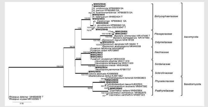
Figure 2 Families of isolated endophyte fungi of four mangrove species. A phylogram obtained from the BI, ML and MP analyses of the ITS dataset. The sequences obtained in this study in bold. The subsequent probabilities ≥ 95 % of BI are represented by thick branches. Bootstrap support values > 70 % are indicated near nodes as MP/ML. * • Bootstrap support values < 70 %. 
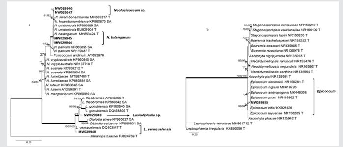
Figure 3 Phylogram of genera in Botryosphaeriaceae. Six of the 17 mangrove isolates are closely grouped with the genera Lasiodiplodia and Neofusicoccum (3a). A phylogram of Didymellaceae genera, indicating that one of the isolates obtained from Rhizophora racemosa (in bold) is grouped with different species of Epicoccum (3b). Phylograms obtained from BI, ML and MP analyses of the ITS dataset. The subsequent probabilities ≥ 95 % of IB are represented by thick branches. Bootstrap support values > 70 % are indicated near nodes as MP/ML. * • Bootstrap support values < 70 %. 
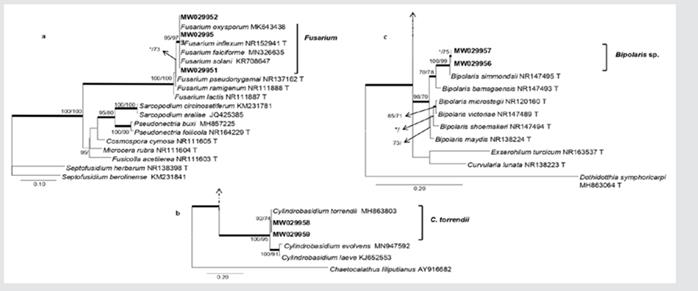
Figure 4 Phylogenetic analysis of genera in Nectriaceae. Mangrove isolates (in bold) are grouped with different species of Fusarium (4a). Phylogram with genus Physalacriaceae, the isolates obtained from mangroves are clearly grouping with Cylindrobasidium torrendii (4b). Phylogram of Pleosporaceae genera. Isolations obtained from Pelliciera rhizophorae (in bold) suggest a close relationship with Bipolaris simmondsii (4c). Phylograms obtained from BI, ML and MP analyses of the ITS dataset. The subsequent probabilities ≥ 95 % of IB are represented by thick branches. Bootstrap support values > 70 % are indicated near nodes as MP/ML. * • Bootstrap support values < 70 %. 

Phylogeny of Physalacriaceae included nine previously described species; two isolates obtained from mangroves (MW029959, MW029958) were closely related to Cylindrobasidium torrendii (Fig. 4b). In Pleosporaceae, 16 species described earlier were included in the phylogenetic analysis; two isolates obtained from mangroves (MW029957, MW029956) formed a separate clade within the family (Fig. 4c). In Psathyrellaceae, 13 species were included in the phylogenetic analyses, of these, a single isolate obtained from mangroves (MW029960) grouped with Coprinellus radians (Fig. 5a). In Sclerotiniaceae, the isolate (MW029950) was closely grouping with two species in the same clade, Ciboria aestivalis and Moellerodiscus lentus (Fig. 5b). In Sordariaceae, a mangrove isolate (MW029954) was grouping with Neurospora crassa, N. pannonica, N. sitophila and N. tetrasperma (Fig. 5c).
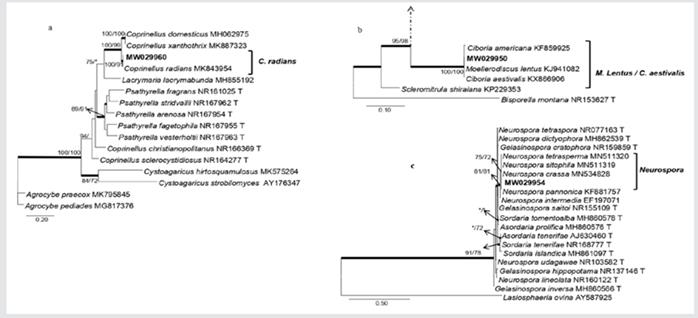
Figure 5 Phylograms obtained from genera in Psathyrellaceae indicating that one of the isolations obtained from Rhizophora racemosa (in bold) is closely related to Coprinellus radians (5a). Phylogram of genera in Sclerotiniaceae indicating that one of the isolates obtained from Laguncularia racemosa (in bold) groups with Moellerodiscus lentus and Ciboria aestivalis (5b). Phylogram obtained from genera in Sordariaceae,shows that isolations obtained from branches of Laguncularia racemosa (in bold) groups with four species of Neurospora (5c). Phylograms obtained from BI, ML and MP analyses of the ITS dataset. The subsequent probabilities ≥ 95 % of IB are represented by thick branches. Bootstrap support values > 70 % are indicated near nodes as MP/ML. * • Bootstrap support values < 70 %. 

DISCUSSION

This study represents the first phylogenetic approach of endophytic fungi associated with mangrove trees in an area of the Colombian Pacific coast. Nine fungal genera were identified based on the DNA sequence data for the ITS region, a gene region widely used as a molecular marker in fungal identification and taxonomic studies (Hibbett et al., 2011; Joel and Bhimba, 2013). Based on the phylogenetic analysis, the identity of the isolates were verified and revealed that the endophytes collected in this study closely grouped with species such as Ciboria aestivalis, Coprinellus radians, Cylindrobasidium torrendii, Epicoccum nigrum, Fusarium oxysporum, Lasiodiplodia theobromae, Lasiodiplodia venezuelensis, Neofusicoccum batangarum and Neurospora crassa, and two isolates that could not be confirmed to species level, Bipolaris sp. and Neofusicoccum sp., all are reported for the first time at the Colombian Pacific coast.

The fungi obtained in this study belong mostly to Ascomycota, with fourteen isolates within seven genera; meanwhile, three isolates were members of Basidiomycota. Interestingly, previous studies have confirmed that the aerial and submerge parts of mangrove trees, are largely dominated by ascomycetes, while basidiomycetes have a lower proportion (Alias et al., 2010; Sun and Guo, 2012; Abdel et al., 2014; Simões et al., 2015), this is consistent with the saprophytic activity that present a large number of Basidiomycetes (Agrios, 2005; Mohapatra, 2008). While most Basidiomycetes are mostly found in terrestrial environments (Shearer et al., 2007), the Ascomycetes are considered as an important ecological group, with a wide variety occurring in diverse environments, including marine environments (Jones and Tan, 1987; Hyde and Jones, 1988; Abdel-Wahab, 2005; Vittal and Sarma, 2006; Alias et al., 2010; Suetrong et al., 2017). Moreover, a wide range of variables including host age, architecture, brightness levels, genotype, height of crown, among others, have been considered to affect fungal community structure, as well as endophyte-plant interactions (Saikkonen, 2007).

Two genera in the Botryosphaeriaceae, including Lasiodiplodia and Neofusicoccum were obtained in this study, it is consistent with previous studies reporting their wide range of ecologically diverse fungi, found in all geographical and climatic areas of the world (except in polar regions) (Slippers and Wingfield, 2007; Phillips et al., 2013). For instance, reports of Botryosphaeria, Lasiodiplodia, and Neofusicoccum in mangroves of Brazil (De Souza et al., 2013), Neofusicoccum in China (Xing et al., 2011) and fourteen species belonging to four genera, Botryosphaeria, Diplodia, Lasiodiplodia, and Neofusicoccum in mangroves of South Africa (Osorio et al., 2017), confirms the ubiquity of the species belonging to this fungal family.

Neofusicoccum was the most commonly isolated genus, given the ability of species of this genus to inhabit a wide variety of plant species and geographical areas worldwide (Slippers et al., 2005; Sakalidis et al., 2011). This is consistent with our results, since Neofusicoccum was the most isolated genus, indicating its wide geographical range (Shetty et al., 2011).

Lasiodiplodia theobromae is considered a highly phytopathogenic fungus (Slippers and Wingfield, 2007), which is commonly found in tropical and subtropical regions (Punithalingam, 1980; Burgess et al., 2006; Phillips et al., 2013; Mehl et al., 2017). Previous reports indicate that this species is associated with different host plants, causing plan diseases and being responsible for serious damage to crops of economic importance (Coutinho et al., 2017). It has also been reported as the causal agent of rot, withering and necrosis of seeds in mangrove plants in India (Misral, 2002), and also as a fungal pathogen of mangrove plants in the Niger Delta in Nigeria (Ukoima and Amakiri 2000), possibly in response to an imbalance in the mangrove ecosystems. It is then of concern, considering that mangroves in in Buenaventura are under constant stress due to the anthropogenic disturbances observed during field work, although no obvious symptoms of disease were found. In this sense, further studies are highly recommended to determine the degree of pathogenicity of this species in Colombian mangroves.

In this study, phylogenetic analyses supported that the isolate MW029948 is closely related to Lasiodiplodia venezuelensis, isolated from branches of R. racemosa. To date, this species has not been widely reported in other parts of the world; L. venezuelensis is considered as a rare species having geographically limited distribution (Mohali, et al., 2017). This is the first report of L. venezuelensis associated with mangrove trees in Colombia.

The isolates MW029951, MW029952 and MW029953 grouped within the genus Fusarium, and subsequent analyses revealed their proximity to F. oxysporum, which is a widely existing species (Munkvold, 2017). This isolate was obtained from three of the four mangroves sampled in San Pedro; L. racemosa, M. oleifera and P. rhizophorae, and was the only isolated species from M. oleifera. The genus includes numerous ubiquitous species that can colonize a wide range of habitats (Munkvold, 2017). In mangrove environments the genus has been found colonizing the mangroves of Mangalvan (Pal and Purkayastha, 1992), the leaves and branches of Avicennia schaueriana, L. racemosa and R. mangle in Brazil (De Souza et al., 2013), and recently in microbiome of the mangroves rhizosphere of Avicennia marina in the Red Sea, Saudi Arabia (Simões et al., 2015). Also, members of the F. oxysporum complex are known to cause vascular withering diseases, and are important as root rot pathogens (Munkvold, 2017). Although F. oxysporum includes both pathogenic and nonpathogenic strains, it will be then important to conduct future studies to investigate its pathogenicity in mangroves, and thus to estimate a possible threat to these ecosystems.

Members of the Didymellaceae are cosmopolitan (Aveskamp et al., 2008, 2009; Chen et al., 2015a, 2015b). The isolate MW029955 resides in this family, being phylogenetically closely related to Epicoccum nigrum (99 / 100 %). Different species of Epicoccum are known to produce bioactive secondary metabolites with antiviral (Guo et al., 2009), antifungals (Harwoko et al., 2019), anti-inflammatory (Gonda et al., 2016), and cytotoxic activities (Wang et al., 2010). Among these species, the E. nigrum species complex (Favaro et al., 2011), has been an important part of research on bioactive compounds of endophytic fungi isolated from the mangrove Acanthus ilicifolius L. (Yan et al., 2019). Such research is also facilitated due to the wide geographical distribution and high hosts’ variability (Perveen et al., 2017; Ahumada et al., 2018; Rhim et al., 2019; El-Sayed etal., 2020).

The current phylogenetic analyses confirmed that the isolate obtained from R. racemosa (MW029960) corresponded to Coprinellus radians. Coprinellus is one of the most important lineages of Coprinoid fungi in the Psathyrellaceae (Hussain et al., 2018), with approximately 80 described species (Kirk et al., 2008; Nagy et al., 2012; Gomes and Wartchow, 2014). These fungi are common saprotrophs in wood chips, leaf blades and herbivorous manure (Schafer, 2010). Coprinellus radians has been mainly isolated from human patients (Lu et al., 2020), however, information on its relationship to mangrove trees still remains very limited. Neurospora crassa isolated from branches of L. racemosa in San Pedro, was also isolated from leaves of Rhizophora mucronata Lam (Joel and Bhimba, 2013) to determine their antimicrobial and cytotoxic activity. Neurospora is common in humid tropical and subtropical regions (Turner et al., 2001), also found in mangrove sediments in India (Thamizhmani and Senthilkumaran, 2012) and in the mycobiome of the rhizosphere of the red Sea marine Avicennia mangroves (Simões et al., 2015). Both C. radians and N. crassa are species that represent a promising source of bioactive molecules with potential use in the pharmaceutical industry (Demers et al., 2018).

Of the four mangrove species sampled in San Pedro, L. racemosa and R. racemosa have previously been reported as hosts of endophytic fungi (Vázquez et al., 2000; De Souza et al., 2013; Ukoima et al., 2013), while for M. oleifera and P. rhizophorae there is still no data available. Likewise, information for Bipolaris sp., Ciboria aestivalis and Cylindrobasidium torrendii known to be associated with mangrove trees remain limited. Regarding M. oleifera and P. rhizophorae, the former belongs to a genus native to lowlands of the rainforests of South America, Central America and the Caribbean islands (Bernal et al., 2015), and the latter is the only species of the genus, being native to Central America from Honduras to Ecuador (Ulloa et al., 2017). This limited distribution of hosts, coupled with the limited fungal studies associated with mangrove ecosystems, and illustrates the lack of information on fungal microorganisms associated with these trees worldwide. Bipolaris (MW029956 and MW029957) includes members considered important pathogens of more than 60 plant genera (Sivanesan, 1984; Agrios, 2005; Manamgoda et al., 2011; Hyde et al., 2014).

Phylogenetic analyses also confirmed that the isolate MW029950 obtained from L. racemosa was closely relate to C. aestivalis. This species has been found in herbarium specimens of Cydonia oblonga Miller (Rosaceae) (Galan and Palmer, 2001). In addition, current analyses supported that the isolates MW029958 and MW029959 were closely related to C. torrendii, a fungus that causes weak and limited decomposition of wood (Floudas et al., 2015). These fungal groups are reported for the first time from asymptomatic branches of mangroves in Colombia.

Despite Colombia’s great floristic richness, studies in terms of its associated mycobiota remain very limited. In this sense, this study contributes to the knowledge of fungal diversity associated with mangrove trees in Colombia.

ACKNOWLEDGMENT

We acknowledge to the National Environmental Licensing Authority (ANLA) for sampling permits (Permission: 2019080900-1) under the National Framework granted by resolution No. 01789., as well as the students from the Biology Program, Universidad del Quindío for assistance in the field. Likewise, thanks to the Agro-Industry and Biology Departments of the Universidad del Quindío for providing the physical spaces to carry out the laboratory procedures.

LITERATURE CITED

Abdel MA, Hodhod MS, Bahkali AH, Jones EB. Marine fungi of Saudi Arabia. Bot Mar. 2014; 57 323-335. https://doi.org/10.1515/bot-2014-0010Links ]

Abdel-Wahab MA. Diversity of marine fungi from Egyptian Red Sea mangroves. Bot Mar. 2005; 48 348-355. https://doi.org/10.1515/BOT.2005.047Links ]

Agrios GN. Plant pathology. 5th ed. London: Elsevier/Academic Press; 2005.948. [ Links ]

Ahumada R, Novoa V, Becerra J. Morphological response to salinity, temperature, and pH changes by marine fungus Epicoccum nigrum. Environ Monit Assess. 2018;191. https://doi.org/10.1007/s10661-018-7166-5Links ]

Akaike H. A new look at the statistical model identification. IEEE Trans Autom Control. 1974;19 716-723. [ Links ]

Alias SA, Zainuddin N, Jones EB. Biodiversity of marine fungi in Malaysian mangroves. Bot Mar. 2010;53 545-54. https://doi.org/10.1515/bot.2010.066Links ]

Altschul SF, Gish W, Miller W, Myers EW, Lipman DJ. Basic local alignment search tool. J Mol Biol. 1990;21 5403-10. https://doi.org/10.1016/S0022-2836(05)80360-2Links ]

Álvarez L. R. Los manglares de Colombia y la recuperación de sus áreas degradadas: revisión bibliográfica y nuevas experiencias. Mad Bosq. 2003;93-25. https://doi.org/10.21829/myb.2003.911286Links ]

Arnold AE. Understanding the diversity of foliar endophytic fungi: progress, challenges, and frontiers. Fungal Biol Rev. 2007;2151-66. https://doi.org/10.1016/j.fbr.2007.05.003Links ]

Aveskamp MM, De Gruyter J, Crous PW. Biology and recent developments in the systematics of Phoma, a complex genus of major quarantine significance. Fungal Divers. 2008;311-18 [ Links ]

Aveskamp MM, Verkley G, De Gruyter J, Murace MA, Perelló A, Woudenberg JHC, Groenewald JZ, Crous PW. DNA phylogeny reveals polyphyly of Phoma section Peyronellaea and multiple taxonomic novelties. Mycologia. 2009;101 363-82. https://doi.org/10.3852/08-199Links ]

Bernal R, Gradstein SR, Celis M. Catálogo de plantas y líquenes de Colombia, Mora oleifera (Hemsl.) Ducke . Bogotá: Inst Cienc Nat, Univ Nal Col; 2015. http://catalogoplantasdecolombia.unal.edu.coLinks ]

Bolívar-Anillo HJ, Visbal AZ, Serrano MC, Moreno HS, Daza DAV. A preliminary review on the Importance of Colombian mangroves as a source of endophytic microorganisms relevant in pharmaceutical industry. J Trad Chin Med. 2020;3006 [ Links ]

Burgess TI, Barber PA, Mohali S, Pegg G, de Beer W, Wingfield MJ. Three new Lasiodiplodia spp. from the tropics, recognized based on DNA sequence comparisons and morphology. Mycologia. 2006;98 423-35. https://doi.org/10.1080/15572536.2006.11832677Links ]

Chen Q, Jiang JR, Zhang GZ, Cai L, Crous PW. Resolving the Phoma enigma. Stud Mycol. 2015;82 137-217 [ Links ]

Chen Q, Zhang K, Zhang GZ, Cai L. A polyphasic approach to characterise two novel species of Phoma (Didymellaceae) from China. Phytotaxa. 2015;197 267-81. https://doi.org/10.11646/phytotaxa.197.4.4Links ]

Coutinho I, Freire F, Lima C, Lima J, Gonçalves F, Machado A, Silva A, Cardoso J. Diversity of genus Lasiodiplodia associated with perennial tropical fruit plants in northeastern Brazil. Plant Pathol. 2017;66 90-104. https://doi.org/10.1111/ppa.12565Links ]

Cornejo X, Peña E, Cantera J, Silverstone P, Linares R, Monzón M, Bonifaz C. Mangrove forest of the Pacific coast of Colombia: 12-20. In: Plants of South American pacific mangrove swamps (Colombia, Ecuador, Peru) [ Links ]

Darriba D, Taboada G, Doallo R, Posada D. jModelTest2: more models, new heuristics and parallel computing. Nat Meth. 2012;9. https://doi.org/10.1038/nmeth.2109Links ]

De Souza SFL, Dumaresq ASR, Lacava PT, Harakava R, Azevedo JL, de Melo IS, Pizzirani A. Species diversity of culturable endophytic fungi from Brazilian mangrove forests. Curr Gen. 2013;59 153-66. [ Links ]

Demers DH, Knestrick MA, Fleeman R, Tawfik R, Azhari A, et al. Exploitation of mangrove endophytic fungi for infectious disease drug discovery. Mar Drugs. 2018;16. https://doi.org/10.3390/md16100376Links ]

El-Sayed ER, Ahmed AS, Abdel-hakim HK. A novel source of the cardiac glycoside digoxin from the endophytic fungus Epicoccum nigrum: Isolation, characterization, production enhancement by gamma irradiation mutagenesis and anticancer activity evaluation. J Appl Microbiol. 2020;128 747-62. https://doi.org/10.1111/jam.14510Links ]

Favaro LC, De Melo FL, Aguilar CI, Araújo WL. Polyphasic analysis of intraspecific diversity in Epicoccum nigrum warrants reclassification into separate species. PLoS One. 2011;6. https://doi.org/10.1371/journal.pone.0014828Links ]

Floudas D, Held BW, Riley R, Nagy LG, Koehler G, Ransdell AS, Younus H, Chow J, Chiniquy J, Lipzen A, Tritt A, Sun H, Haridas S, LaButti K, Ohm RA, Kües U, Blanchette RA, Grigoriev IV, Minto RE, Hibbett DS. Evolution of novel wood decay mechanisms in Agaricales revealed by the genome sequences of Fistulina hepatica and Cylindrobasidium torrendii. Fungal Gen Biol. 2015;76 78-92 [ Links ]

Galan R, Palmer JT. The occurrence of the rare Ciboria aestivalis in Europe. Czech Mycol. 2001;52 227-87. https://doi.org/10.33585/cmy.52404Links ]

Gamboa-Gaitán MA, Otero-Ospina JT. Colombian vanilla and its microbiota. III. Diversity and structure of the endophytic community. Acta Bot Hung. 2016;58 241-56. https://doi.org/10.1556/abot.58.2016.3-4.2Links ]

García C. Diagnóstico de las áreas marinas y costeras protegidas, y de las áreas de manejo en el Pacífico colombiano: 65. Fundación MarViva, Colombia; 2010. https://www.researchgate.net/publication/323656733Links ]

Gilbert GS, Mejia M, Rojas E. Fungal diversity and plant disease in mangrove forests: salt excretion as a possible defense mechanism. Oecologia. 2002;132 278-85. https://doi.org/10.1007/s00442-002-0966-9Links ]

Giri C, Ochieng E, Tieszen LL, Zhu Z, Singh A, Loveland T, Masek J, Duke N. Status and distribution of mangrove forests of the world using earth observation satellite data. Global Ecol Biogeogr. 2011;20 154-59 [ Links ]

Gomes ARP, Wartchow F. Coprinellus arenicola, a new species from Paraíba, Brazil. Sydowia. 2014;66 249-56. https://doi.org/10.12905/0380Links ]

Gonda S, Kiss-Szikszai A, Szucs Z, Balla B, Vasas G. Efficient biotransformation of non-steroid anti-inflammatory drugs by endophytic and epiphytic fungi from dried leaves of a medicinal plant, Plantago lanceolata L. Int Biodeter Biodegrad. 2016;108 115-21. https://doi.org/10.1016/j.ibiod.2015.12.018Links ]

Guindon S, Gascuel O. A simple, fast and accurate method to estimate large phylogenies by maximum-likelihood. Syst Biol. 2003;52 696-704. https://doi.org/10.1080/10635150390235520Links ]

Guo H, Sun B, Gao H, Chen X, Liu S, Yao X, Liu X, Che Y. Diketopiperazines from the Cordyceps-colonizing fungus Epicoccum nigrum. J Nat Prod. 2009;722 115-9. https://doi.org/10.1021/np900654aLinks ]

Harwoko H, Daletos G, Stuhldreier F, Lee J, Wesselborg S, Feldbrügge M, Müller WEG, Kalscheuer R, Ancheeva E, Proksch P. Dithiodiketopiperazine derivatives from endophytic fungi Trichoderma harzianum and Epicoccum nigrum. Nat Prod Res. 2019;181-9. https://doi.org/10.1080/14786419.2019.1627348Links ]

Hibbett DS, Ohman A, Glotzer D, Nuhn M, Kirk P, Nilsson RH. Progress in molecular and morphological taxon discovery in fungi and options for formal classification of environmental sequences. Fungal Biol Rev. 2011;25 38-47 [ Links ]

Hussain S, Usman M, Afshan N-ul-S, Ahmad H, Khan J, Khalid AN. The genus Coprinellus (Basidiomycota; Agaricales) in Pakistan with the description of four new species. MycoKeys. 2018;39 41-61. https://doi.org/10.3897/mycokeys.39.26743Links ]

Hyde KD, Jones EBG. Marine mangrove fungi. PSZNI Mar Ecol. 1988;915-33. https://doi.org/10.1111/j.1439-0485.1988.tb00196.xLinks ]

Hyde KD, Lee SY. Ecology of mangrove fungi and their role in nutrient cycling - what gaps occur in our knowledge. Hydrobiologia. 1995;295 107-18. https://doi.org/10.1007/BF00029117Links ]

Hyde KD, Nilsson RH, Alias SA, Ariyawansa HA, Blair JE, Cai L, de Cock AWAM, Dissanayake AJ, Glockling SL, Goonasekara ID, Gorczak M, Hahn M, Jayawardena RS, Kan JAL, Laurence MH, Lévesque CA, Li X, Liu JK, Maharachchikumbura SSN, Manamgoda DS, Martin FN, McKenzie EHC, McTaggart AR, Mortimer PE, Nair PVR, Pawłowska J, Rintoul TL, Shivas RG, Spies CFJ, Summerell BA, Taylo PWJ, Terhem RB, Udayanga D, Vaghefi N, Walther G, Wilk M, Wrzosek M, Xu JC, Yan JY, Zhou N. One stop shop: backbones trees for important phytopathogenic 5 genera: I. Fungal Divers. 2014;67 21-125. https://doi.org/10.1007/s13225-014-0298-1Links ]

Joel EL, Bhimba BV. Biological activity of secondary metabolites isolated from mangrove fungi Neurospora crassa. J Environ Biol. 2013;347 29-32 [ Links ]

Jones EBG, Tan TK. Observations on manglicolous fungi from Malaysia. Trans Br Mycol. 1987;89 390-2. [ Links ]

Katoh K, Rozewicki J, Yamada KD. MAFFT online service: multiple sequence alignment, interactive sequence choice and visualization. Brief Bioinf. 2019;201 160-6. https://doi.org/10.1093/bib/bbx108Links ]

Kirk PM, Cannon PF, Minter DW, Stalpers JA. Dictionary of the Fungi. 10th edn. Wallingford: CABI; 2008 [ Links ]

Kumar S, Stecher G, Li M, Knyaz C, Tamura K. MEGA X: Molecular Evolutionary Genetics Analysis across computing platforms. Mol Biol Evol. 2018;351 547-9. https://doi.org/10.1093/molbev/msy096Links ]

Kuraku S, Zmasek CM, Nishimura O, Katoh K. Leaves facilitates on-demand exploration of metazoan gene family trees on MAFFT sequence alignment server with enhanced interactivity. Nucl Acids. 2013;41 W22-8. https://doi.org/10.1093/nar/gkt389Links ]

López A, Roberts C, Tilley A, Hawkins J, Cooke R. Mangroves and people: Lessons from a history of use and abuse in four Latin American countries. For Ecol Manag. 2016;368 151-62 [ Links ]

Lu X, Wang X, Zhang L, Li X, Qi X. Rare fungal keratitis caused by Coprinellus radians. Mycopathologia. 2020;183 89-94. https://doi.org/10.1007/s11046-019-00414-yLinks ]

Manamgoda DS, Cai L, Bahkali AH, Chukeatirote E, Hyde KD. Cochliobolus: an overview and current status of species. Fungal Divers. 2011;513-42. https://doi.org/10.1007/s13225-011-0139-4Links ]

Mejía Q, Molina J, Sanjuan M, Grijalba B, Niño M. Bosque de manglar, un ecosistema que debemos cuidar: 27. Cartagena DT: Univ Jorge Tadeo Lozano, Inst Col Desarr Rur; 2014. [ Links ]

Mehl J, Wingfield MJ, Roux J, Slippers B. Invasive everywhere? Phylogeographic analysis of the globally distributed tree pathogen Lasiodiplodia theobromae. Forests. 2017;8. https://doi.org/10.3390/f8050145Links ]

Misral DS. Development of mixed formation of fungal (Trichoderma) and bacterial (Pseudomonas) biocontrol agents for management of plant disease. Ph.D. Thesis. Univ Agriculture Technology Pantnager; 2002. 185 p. [ Links ]

Mohali SR, Castro F, Úrbez JR, Gubler WD. First report of Lasiodiplodia theobromae and L. venezuelensis associated with blue stain on Ficus insipida wood from the natural forest of Venezuela. For Path. 2017; 47 1-5 https://doi.org/10.1111/efp.1235Links ]

Mohapatra . Textbook of environmental microbiology. New Delhi: I K International Publishing; 2008. 546p [ Links ]

Munkvold GP. Fusarium species and their associated mycotoxins. Meth Mol Biol. 2017;154 251-106. https://doi.org/10.1007/978-1-4939-6707-0_4Links ]

Nagy LG, Kocsubé S, Csanádi Z, Kovács GM, Petkovits T, Vágvölgyi C, Papp T. Re-mind the gap! Insertion-deletion data reveal neglected phylogenetic potential of the nuclear ribosomal Internal Transcribed Spacer (ITS) of Fungi. PLoS One. 2012;7. https://doi.org/10.1371/journal.pone.0049794Links ]

Ordóez NF, Otero JT, Díez MC. Hongos endófitos de orquídeas y su efecto sobre el crecimiento en Vanilla planifolia Andrews. Acta Agron. 2012;61 282-290 [ Links ]

Pal AK, Purkayastha RP. New parasitic fungi from Indian mangrove. J Mycopathol Res. 1992;30 173-176 [ Links ]

Palacios ML, Cantera JR. Mangrove timber use as an ecosystem service in the Colombian Pacific. Hydrobiologia. 2017;803 345-358. https://doi.org/10.1007/s10750-017-3309-xLinks ]

Perveen I, Raza MA, Iqbal T, Naz I, Sehar S, Ahmed S. Isolation of anticancer and antimicrobial metabolites from Epicoccum nigrum; endophyte of Ferula sumbul. Microb Pathog. 2017;110 214-224. https://doi.org/10.1016/j.micpath.2017.06.033Links ]

Petrini O. Fungal endophytes of tree leaves. In: Andrews JH, Hirano SS (Eds). Microbial ecology of leaves. New York, NY: Springer; 1991. https://doi.org/10.1007/978-1-4612-3168-4_9Links ]

Phillips AJL, Alves A, Abdollahzadeh J, Slippers B, Wingfield MJ, Groenewald JZ, Crous PW. The Botryosphaeriaceae: genera and species known from culture. Stud Mycol. 2013;7 651-167 [ Links ]

Prahl Hv. Manglares de Colombia. Villegas, Banco Occidente; 1989. 205p [ Links ]

Punithalingam E. Plant diseases attributed to Botryodiplodia theobromae Pat.. Vadus, Germany; 1980. 121p [ Links ]

Raeder U, Broda P. Rapid preparation of DNA from filamentous fungi. Lett Appl Microbiol. 1985;117-20. https://doi.org/10.1111/j.1472-765X.1985.tb01479.xLinks ]

Rambaut A, Drummond AJ, Xie D, Baele G, Suchard MA. Posterior summarization in Bayesian phylogenetics using Tracer 1.7. Syst Biol. 2018;67 901-904. https://doi.org/10.1093/sysbio/syy032Links ]

Rhim H, Park JY, Lee DJ, Han JI. Epicoccum nigrum-induced respiratory infection in a wild Eurasian scops owl (Otus scops). J Vet Med. 2019;81 1348-1350. https://dx.doi.org/10.1292/jvms.19-0172Links ]

Rineau F, Shah F, Smits MM, Persson P, Johansson T, Carleer R, Troein C, Tunlid A. Carbon availability triggers the decomposition of plant litter and assimilation of nitrogen by an ectomycorrhizal fungus. ISME J. 2013;7 [ Links ]

Ronquist F, Teslenko M, van der Mark P, Ayres DL, Darling A, Höhna S, Larget B, Liu L, Suchard MA, Huelsenbeck JP. MRBAYES 3.2: Efficient Bayesian phylogenetic inference and model selection across a large model space. Syst Biol. 2012;61 539-542. https://doi.org/10.1093/sysbio/sys029Links ]

Sakalidis ML, Hardy GESJ, Burgess TI. Class III endophytes, clandestine movement amongst hosts and habitats and their potential for disease; a focus on Neofusicoccum australe. Australas Plant Path. 2011;40 510-521. https://doi.org/10.1007/s13313-011-0077-3Links ]

Saikkonen K. Forest structure and fungal endophytes. Fungal Biol Rev. 2007;21 67-74. https://doi.org/10.1016/j.fbr.2007.05.001Links ]

Salazar CS, De García MCC. Aislamiento de hongos endófitos en rosa (Rosa hybrida) en Bogotá, Colombia. Rev Iberoam Micol. 2005;22 99-101. https://doi.org/10.1016/S1130-1406(05)70016-4Links ]

Schafer DJ. Keys to sections of Parasola, Coprinellus, Coprinopsis and Coprinus in Britain. Field Mycol. 2010;11 44-51. [ Links ]

Shearer CA, Descals E, Kohlmeyer B, Kohlmeyer J, Marvanová L, Padgett D, Porter D, Raja HA, Schmit JP, Thorton HA, Voglymayr H. Fungal biodiversity in aquatic habitats. Biodivers Cons. 2007;16 49-67. https://doi.org/10.1007/s10531-006-9120-zLinks ]

Shetty KG, Minnis AM, Rossman AY, Jayachandran K. The Brazilian peppertree seed-borne pathogen, Neofusicoccum batangarum, a potential biocontrol agent. Biol Control. 2011;56 91-97. https://doi.org/10.1016/j.biocontrol.2010.09.016Links ]

Simões MF, Antunes A, Ottoni CA, Amini MS, Alam I, Alzubaidy H, Mokhtar NA, Archer JAC, Bajic VB. Soil and rhizosphere associated fungi in gray mangroves (Avicennia marina) from the Red Sea: a metagenomic approach. Genom Proteom Bioinform. 2015;13 310-320. https://doi.org/10.1016/j.gpb.2015.07.002Links ]

Sivanesan A. The bitunicate ascomycetes and their anamorphs. Vaduz, Liechtenstein: J Cramer; 1984. 701p. [ Links ]

Slippers B, Wingfield MJ. Botryosphaeriaceae as endophytes and latent pathogens of woody plants: diversity, ecology and impact. Fungal Biol Rev. 2007;2 190-106. [ Links ]

Slippers B, Summerell BA, Crous PW, Coutinho TA, Wingfield BD, Wingfield MJ. Preliminary studies on Botryosphaeria species from Southern Hemisphere conifers in Australasia and South Africa. Australas Plant Pathol. 2005;34 213-220. https://doi.org/10.1071/AP05020Links ]

Spalding M, Kainuma M, Collins L. World atlas of mangroves. London: Earthscan; 2010. 336 p. [ Links ]

Suetrong S, Preedanon S, Klaysuban A, Gundool W, Unagul P, Sakayaroj J, Promchu W, Sangtiean T. Distribution and occurrence of manglicolous marine fungi from eastern and southern Thailand. Bot Mar. 2017;60 503-514. https://doi.org/10.1515/bot-2016-0107Links ]

Sun X, Guo LD. Endophytic fungal diversity: review of traditional and molecular techniques. Mycology. 2012;3 65-76. [ Links ]

Swofford DL. PAUP*: phylogenetic analysis using parsimony (*and other methods). Sunderland, Massachusetts: Sinauer Associates; 2003. [ Links ]

Thamizhmani R, Senthilkumaran R. Diversity of fungi in selected mangroves along the east coast of India. Int J Curr Microbiol Appl. 2012;129-33 [ Links ]

Thatoi H, Behera BC, Mishra RR, Dutta SK. Biodiversity and biotechnological potential of microorganisms from mangrove ecosystems: a review. Ann Microbiol. 2013;6 31-19. https://doi.org/10.1007/s13213-012-0442-7Links ]

Tomlinson P. The botany of mangroves. 2nd ed. Cambridge: Cambridge Univ; 2016. 432 p [ Links ]

Turner BC, Perkins DD, Fairfield A. Neurospora from natural populations: a global study. Fungal Genet Biol. 2001;32 67-92. https://doi.org/10.1006/fgbi.2001.1247Links ]

Ukoima HN, Amakiri MA. Fungi associated with the roots of Rhizophora mangle, Rhizophora harrisonii, and Avicennia Africana in Port Harcourt Mangrove swamp, Rivers State. Ecol Environ Cons. 2000;13 32-42 [ Links ]

Ukoima HN, Ikata M, Pepple GA. Control of Lasiodiplodia theobromae (PAT) on Rhizophora racemosa using plants extracts. Am J Biotechnol Mol Sci. 2013;31-7 [ Links ]

Ulloa C, Acevedo P, Beck S, Belgrano MJ, Bernal R, Berry PE, Brako L, Celis M, Davidse G, Forzza RC, Gradstein SR, Hokche O, León B, León S, Magill RE, Neill DA, Nee MH, Raven PH, Stimmel H, Strong MT, Villaseñor JL, Zarucchi JL, Zuloaga FO, Jørgensen MP. An integrated assessment of vascular plants species of the Americas. Science. 2017;358 1614-1617. https://doi.org/10.1126/science.aao0398Links ]

Vázquez P, Holguin G, Puerte ME, López-Cortés A, Bashan Y. Phosphate-solubilising microorganisms associated with the rhizosphere of mangroves in a semiarid Coastal lagoon. Biol Fertil Soils. 2000;304 60-46. https://doi.org/10.1007/s003740050024Links ]

Vittal BPR, Sarma VV. Diversity and ecology of fungi on mangroves of bay of Bengal region - an overview. Indian J Mar. 2006;35 308-317 [ Links ]

Wang JM, Ding GZ, Fang L, Dai JG, Yu SS, Wang YH, Chen XG, Ma SG, Qu J, Xu S, Du D. Thiodiketopiperazines produced by the endophytic fungus Epicoccum nigrum. J Nat Prod. 2010;73 1240-1249. https://doi.org/10.1021/np1000895Links ]

White TJ, Bruns T, Lee S, Taylor J. Amplification and direct sequencing of fungal ribosomal RNA genes for phylogenetics: 315-322. In: Innis MA, Gelfand DH, Sninsky JJ, White TJ (Eds). PCR protocols: a guide to methods and applications. San Diego: Academic; 1990 [ Links ]

Wilson D. Endophyte: The evolution of a term, and clarification of its use and definition. Oikos. 1995;73 272-276. https://doi.org/10.1016/B978-0-12-372180-8.50042-1Links ]

Xing XK, Chen J, Xu MJ, Lin WH, Guo SX. Fungal endophytes associated with Sonneratia (Sonneratiaceae) mangrove plants on the south coast of China. For Pathol. 2011;41 334-340. https://doi.org/10.1111/j.1439-0329.2010.00683.xLinks ]

Yan Z, Wen S, Ding M, Guo H, Huang C, Zhu X, Huang J, She Z, Long Y. The purification, characterization, and biological activity of new polyketides from mangrove-derived endophytic fungus Epicoccum nigrum SCNU-F0002. Mar Drugs. 2019;17. https://doi.org/10.3390/md17070414Links ]

Received: July 09, 2022; Accepted: January 30, 2023

*Corresponding autor: osorio.romero17@gmail.com

Creative Commons License This is an open-access article distributed under the terms of the Creative Commons Attribution License