INTRODUCTION
Rocks and minerals, as natural materials, show a wide degree of variation in their physical properties, and measuring some of these properties often serves as a way for rapid and/or precise characterization (Hoyos-Palacio et al., 2015; Filipussi et al., 2019; Ferrari & Gómez 2019).
Rocks can record some of the characteristics of the existing magnetic field at the time of their formation. An important field of geology, paleomagnetism, aims to determine the natural remanent magnetization (NRM) of rocks and interpret it in terms of significant geological events (Butler, 1992; Dunlop & Özdemir, 1997; Valencio, 1980).
Rock magnetism is a field that studies the behavior and magnetic properties of different types of rocks and minerals found in nature. The magnetic characterization of a sample requires measuring various parameters such as magnetic susceptibility, magnetic hysteresis cycles, isothermal/anhysteretic remanence acquisition curves, among others. All these parameters provide essential information to identify the various ferromagnetic minerals present in a sample and their magnetic state (superparamagnetic, single-domain, pseudo-single domain, multidomain), which mainly depends on the size and shape of the grains or crystals (Dunlop & Özdemir, 1997; Ivanchenko et al., 2012).
Rock magnetic studies are usually carried out in addition to any paleomagnetic study, with the purpose of determining what ferromagnetic minerals are carrying the NRM, which can have important implications regarding its origin, stability, and history of acquisition.
In addition, knowing the microphysics of minerals constitutes an interesting field of research. It is also a fundamental tool for environmental or paleoenvironmental magnetism, which attempts to characterize the magnetic phases present in recent or ancient sediments and their variation, both spatial and temporal. Moreover, the connection between environmental or paleoenvironmental parameters and the presence and state of one or more ferromagnetic minerals allows extracting important information (Chaparro-Marcos et al., 2014).
Magnetism in rocks is carried by a few minerals, the most important of which are iron and titanium oxides, such as magnetite and hematite (Stacey & Banerjee, 1974; Dunlop & Özdemir, 1997).
Destructive and non-destructive magnetic measurements are taken in the different rock magnetic studies. Magnetic Barkhausen Noise (MBN) is a non-destructive testing technique that is widely used for the microstructural characterization of magnetic materials. This technique is mainly based on the irreversible movements of the walls of the magnetic domains that must overcome pinning points such as grain edges, dislocations, impurities, etc. These imperfections impede the movement of the domain walls until an increase in the applied magnetic field provides the necessary energy to free the walls from these obstacles. These pinning points produce a sudden jump in magnetization, which causes a flux change called MBN (Jiles, 1991; Stefanita, 2008; Neyra-Astudillo, 2017; Neyra-Astudillo et al., 2018).
In this work, the MBN method is attempted as an alternative technique to study the magnetic characteristics of rock samples that contain different variable proportions of ferromagnetic minerals such as magnetite (Fe3O4). This work evaluates the feasibility of applying the MBN method in the study of magnetic rocks, as there are no references to it in the literature.
METHODOLOGY
Measurements were carried out on 11 cylindrical samples containing different percentages of magnetite. The MBN was measured on the two circular flat faces. The magnetic yoke was fed with a variable current used to generate the excitation field. The variation of the RMS value of the MBN signals was observed in relation to the known magnetic properties of the studied rocks.
Materials
Magnetite (Fe3O4) usually appears in common rocks of the earth's crust as an accessory mineral, that is, in abundances that rarely reach a few percentage points. For this work, samples of ore deposits characterized by a high volume percentage of magnetite were studied, which varied from massive (>50 vol%) to disseminated ore (with concentrations around 5 vol%), belonging to IOCG, BIF, porphyry, and skarns deposits (Table 2). They were cut as cylinders with 23 mm diameter and 20 mm height. The samples were previously demagnetized by applying linearly decreasing alternating magnetic fields (AF). The demagnetization of the NRM was completed with fields of low intensity (about 20-30 mT), which indicates that, in all cases, the magnetite is predominantly in a multidomain state. Table 1 indicates the median destructive field of NRM obtained from AF demagnetization, a measure of the coercive force. Other properties of the specimens such as volume, density, and magnetic susceptibility are also included, which were determined at the Paleomagnetism Laboratory of IGEBA. For illustrative purposes, a column is also included with the approximate estimation of the volume percentage of magnetite, calculated from the value of magnetic susceptibility using the relations of Clark and Emerson (1991), and assuming that magnetite is the only ferromagnetic mineral in the samples. Table 2 indicates the origin of each of the samples along with their geological characterization.
Table 1 Rock magnetic and petrophysical characterization of each sample
| Code | Volume (cm3) | Density (g/cm3) (1) | Bulk susceptibility (SI) (2) | Median destructive field (mT) (3) | Approximate magnetite vol% (4) |
|---|---|---|---|---|---|
| S01 | 10,2 | 2,62 | 0,143 | 11,0 | 4 |
| S02 | 10,5 | 2,72 | 0,255 | 9,5 | 7 |
| S03 | 10,4 | 2,90 | 0,468 | 3,5 | 12 |
| S04 | 10,2 | 3,34 | 0,863 | 2,0 | 21 |
| S05 | 10,3 | 3,11 | 1,379 | 4,5 | 30 |
| S06 | 10,3 | 3,47 | 1,586 | 6,0 | 33 |
| S07 | 10,2 | 3,24 | 2,195 | 1,5 | 42 |
| S08 | 9,7 | 3,92 | 2,654 | 8,5 | 47 |
| S09 | 10,4 | 4,46 | 3,519 | 12,5 | 55 |
| S10 | 9,9 | 4,32 | 4,576 | 16,0 | 63 |
| S11 | 10,2 | 4,63 | 4,874 | 11,0 | 65 |
(1) Determined by immersion method. (2) Measured with Kappabridge MFK1-FA Agico, property of IGEBA (UBA-CONICET), using a field of 200 A/m, corrected by self-demagnetization. (3) Obtained from AF demagnetization curves. (4) Estimated from magnetic susceptibility according toClark and Emerson relations (1991).
Source: Authors
Table 2 Origin of the analyzed samples
| ID | Origin |
|---|---|
| S01 | Stockwork with phyllic alteration, Grasberg porphyry, Indonesia |
| S02 | Lower andesite, Candelaria IOCG, Chile |
| S03 | Banded Iron Formation (BIF), Eyre Peninsula, South Australia |
| S04 | |
| S05 | Stockwork, Grasberg porphyry, Indonesia |
| S06 | Skarn Ertsberg, Grasberg, Indonesia |
| S07 | Banded Iron Formation (BIF), Eyre Peninsula, South Australia |
| S08 | Magnetite breccia in lower andesite (magnetite-chalcopyrite), Candelaria IOCG, Chile |
| S09 | Skarn Doz, Grasberg, Indonesia |
| S10 | Lower andesite with chalcopyrite, Candelaria IOCG, Chile |
| S11 | Lower andesite, Candelaria IOCG, Chile |
Source: Authors
BIF (banded iron formation): chemically precipitated sedimentary rocks, most of them during the Archean, probably coinciding with the oxygenation of the oceans leading to massive precipitation of iron oxides. Most BIFs are highly metamorphosed. IOCG (iron oxide copper gold ore deposits): copper-gold ores within iron oxide dominant gangue. They are metasomatic rocks formed due to large alteration events driven by intrusive activity. Skarn: metasomatic rocks formed by the replacement of carbonate rocks caused by contact with an intrusive body. Stockwork: a system of veins developed in the core of porphyry copper-gold deposits in response to high-temperature hydrothermal alterations in calc-alkaline intrusions.
MBN measurement system
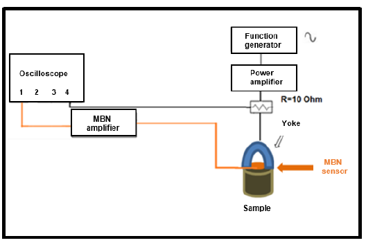
The diagram in Figure 1 shows the scheme of the measurement system. The tests were carried out using a magnetic yoke as an excitation source. This yoke was placed on each of the flat faces of the samples. The magnetic sensor coil was held between the magnetic yoke arms. The excitation magnetic field was achieved with a 10 Hz sinusoidal current produced by a Stanford function generator with a maximum voltage of 8 V, which was then amplified. The excitation and detection procedures were performed on the two circular faces of the cylindrical specimen, and then both results were averaged. Measurements were made with a four-channel digital oscilloscope (LeCroy Wave Runner 44MXi). Digitization was performed with a sampling frequency of 5 Ms/s, repeating each measurement five times on each face and considering their averages. In this way, the current on the yoke, the MBN signals, and the excitation voltage were recorded in three channels of the oscilloscope. Figure 2a shows the photograph of the measurement system. In Figure 2b, the magnetic probe (yoke and sensor coil) is positioned over a sample.

Source: Authors
Figure2. a) Photograph of the measurement system, b) magnetic yoke and sensor coil positioned over the specimen
To study the MBN signals, they were first digitally filtered in order to reduce the influence of noise. To this effect, a 4th-order Butterworth-type band pass filter in the range between 5 and 200 kHz was applied to the signals. Then, the RMS (root mean square) value of each filtered signal was calculated, and the corresponding averages were determined.
RESULTS AND DISCUSSION
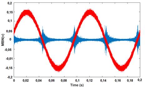
Figure 3 shows the MBN signals (blue) for specimen S10 after they were filtered. They were obtained by exciting the yoke (red). From the filtered signals, the RMS value of the MBN was calculated for two complete cycles of excitation.
In Figure 4, the average of the RMS values evaluated in both opposite flat faces of the samples was plotted as a function of the bulk magnetic susceptibility for all the specimens. As magnetic susceptibility depends on the amount of ferromagnetic minerals present in the sample, the slight increasing trend observed in the figure indicates that the greater the amount of magnetite, the more intense the RMS of the MBN measured. This is particularly true for samples with lower magnetite contents, since the magnetite surfaces in contact with the sensor are smaller. However, for samples with more than, say, 20 vol% magnetite, the effect of concentration becomes less significant, and the correlation worsens.

Source: Authors
Figure 4 Graph of the RMS values as a function of the bulk magnetic susceptibility for all specimens (only the numbers in the rock IDs are shown)
In Figure 5, the RMS value is plotted for each test piece as a function of the median destructive magnetic field (shown in Table 1), which is considered to be indicative of the coercive force of the material. For multidomain materials, the coercive force increases with the presence of greater domain wall pinning and dislocations. Samples with lower magnetic susceptibility (S01 and S02) are clear outliers of any trend. For these samples, the very low values of the measured RMS are due to the low magnetite concentration, as previously discussed. For the rest of the samples, there is a linear relationship between the RMS value and the coercive force, with a positive correlation (R2=0,605). In particular, the BIF samples are the ones with the lowest RMS and coercive force, which is possibly due to recrystallization effect experienced by these samples (S03, S04, S07) during metamorphism, which leads to annealing with the consequent decrease in wall pinning. At the other extreme, magnetites of metasomatic origin (S06, S08, S09, S10, S11) have higher values of coercivity and RMS, possible evidence of the effect of impurities and/or stress.

Source: Authors
Figure 5 Graph of the RMS values as a function of median destructive field of the natural remanent magnetization (only the numbers of the rocks are showed). Open symbols are used for outliers (samples with low RMS due to low magnetite concentration).
Other mechanisms could be studied to explain the fluctuation in the RMS value. This is an initial approach, and those relationships will be evaluated in future works.
CONCLUSION
This was the first time in Argentina that samples of rocks with different origins and varying proportions of magnetite were studied via the MBN technique.
The linear adjustment of the RMS values of the MBN with respect to the percentage of ferromagnetic minerals and magnetic susceptibility showed an increasing trend. A positive correlation was observed between the coercive force (estimated from the median destructive field) and the RMS of the MBN. The researchers interpret that the technique is sensitive to the abundance of magnetic material and that, for massive magnetite samples, it is sensitive to coercivity. In turn, this is conditioned by the geological history of the sample: metamorphosed rocks have lower RMS values, whereas rocks of metasomatic origin (particularly skarns and IOCG) have higher RMS values and coercive force.


