


 English (pdf)
 Article in xml format
 Article references
 Send this article by e-mail
 Cited by SciELO
  Cited by Google
  Similars in
SciELO
Similars in
SciELO  Similars in Google
 Permalink
INTRODUCTION
The ear, responsible for hearing, as the other organs, is subject to pathological changes, such as otitis 1. Such diseases, seen often in clinical veterinary routine, causing great discomfort to dogs, and is common in breeds that have ears drooping, ears with congenitally stenosis of the pinna, dogs with atopic dermatitis and in dogs that live in hot, humid climates 2.
According to Oliveira et al 3, and Gregorio 4, the ear of the healthy dog has a microbiology composed by coccus, bacilli and yeasts that can be found in dogs otopatas, where, in this case, the microbiota will be changed. The diagnosis of otitis externa is based on anamnesis and confirmed by clinical examination, along with exams cytologic, microbiological, diagnostic imaging and video otoscopy 3.
According to Gregorio 4 the antibiogram is indicated in any case that there is suppurative otitis media, chronic relapse and in cases which shall be identified to the cytological examination the presence of bacilli. The treatment commonly performed consists in the elimination of primary causes, cleaning and drying the ears and, especially, the interruption of the infection 2.
The tolerance to antimicrobial agents increase the demand for natural therapies to treat this problem 5,6. Products such as phytotherapy and the use of honey from bees of the genus Apis are widely used as an alternative to conventional therapies 7,8.
The products of the stingless bees have medicinal properties such as antibacterial, antifungal effect, antitumor and healing but are still little known, and may be a lower cost alternative for the treatment of external otitis and. According to Vit 9, honey of these bees have been shown to be an alternative in the treatment of otitis media by assisting the healing of diseases dermatology. In accordance with Bobány et al 10, honey of the stingless bee Tetragonisca angustula showed clear antimicrobial action against mixed cropping of bacilli, coccus and yeasts from auditory meatus of dogs otopatas. The bees of the genus Melipona has been the subject of studies that have shown their effectiveness against various pathogenic bacteria 6.
The geopropolis is a product of the stingless bees composed by a mixture of wax, resin, vegetable fibers and earth or clay, with a function for external structures (tubes and inputs) and internal (honeycombs creates, cover casing and pots of food) 11,12. Cunha 13, working with ethanolic extract of geoprópolis (EEGP) in hexane fractions (FH), chloroform (FC) and ethyl-acetate (FAc), by scanning electron microscopy (SEM) found results that suggest that geopropolis of Melipona scutellaris is a promising source of active compounds against some bacteria, with greater cytotoxicity for tumor cells to normal and also capable of acting on biofilms of Streptococcus mutans, and may be useful in controlling diseases dependent biofilm, related to this microorganism.
Campos et al 14 working with Melipona orbignyi, found in Mato Grosso do Sul, Brazil, indicate that the geopropolis of this species has antimicrobial activity and potential to help combat oxidative stress and the proliferation of tumor cells.
Araújo et al 15, working with Melipona fasciculata, found an antimicrobial effect promising geopropolis in combination with chloramphenicol due to bacterial resistance to antibiotics.
The objective of this study was to test the antibacterial property of geopropolis bee Mandaçaia (Melipona quadrifasciata) in order to prove the efficacy of this product forward to micro-organisms identified in the external auditory meatus of dogs otopatas, clinically diagnosed with otitis externa in the Clinic School of Veterinary Medicine of Unifeso.
MATERIALS AND METHODS
Study location. The experiment was conducted on campus Quinta do Paraiso of Centro Universitário Serra dos Órgãos (Unifeso) in two places: Clinic School of Veterinary Medicine and Microbiology lab.
The campus of Unifeso is located in the state of Rio de Janeiro in the Southeast Region of Brazil, at the geographic position: Latitud 22º 24’ 44” S, Longitud 42º 57’ 56” W and an altitude of 871m.
Preparation of samples of the ears. Secretions from ear of four dogs, randomly selected, clinically diagnosed with otitis externa were obtained by means of sterile swabs. The four dogs were never treated otitis.
Of this material was prepared as blades for staining by Gram stain and identification of agents, observing, during the procedure, the practice of sterilization.
After that, the swabs were immersed in test tubes containing Agar Brain Heart Infusion (BHI - 1.85 g in 50ml) and incubated at 37°C for 24 hours were made slides in order to confirm the growth of bacteria by the method of Gram staining.
Obtainment of the geopropolis. Approximately 40 grams of propolis collected from hives of bees of the species Melipona quadrifasciata were obtained in the apiary Serrano / Teresópolis- RJ, and stored in a plastic bag for later alcoholic extraction, following the methodology developed by Park, Ikegaki 16.
Preparation of the alcoholic extract of geopropolis. The geopropolis was crushed in a mortar with the aid of the pistil and after, was weighed 5 aliquots of 0.8g of geopropolis placed in tubes of trials and adding 10 ml of alcoholic’s diluent in different concentrations:50%, 60%, 70%, 80%, 90%, with intention of verifying the possible efficiency of geopropolis of Melipona quadrifaciata in lower concentrations than that (80%) recommended by by Park, Ikegaki 16 as a dilution more efficiently.
Made the dilution, the test tubes were placed in agitator and was then performed the extraction of the active principle, putting the tubes in a water bath in agitation at 70°C for 30 minutes.
After this step, the tubes removed from the water bath were brought into a centrifuge for 10 minutes and was subsequently initiated the withdrawal of the liquid supernatant from each tube for glass amber corresponding to each dilution and stored in refrigerator.
Antibiogram with alcoholic extract of geopropolis. The samples of the ear secretions incubated in the midst BHI, was collected an aliquot of each tube, with automatic pipette 10µL, placed in a test tube containing the BHI so doing a pool of bacteria. Were made 10 test tubes (two tubes for each concentration of 50% 60% 70% 805 90%) and, at each concentration, used 0.5ml and 1 ml of extract of geoprópolis respectively. The tubes were taken to oven at 37°C for 24 hours.
After reading the results of the antibiogram, for confirmation of the antibacterial action of geopropolis, was prepared blades for staining by Gram stain for identification of possible microbial agents, in accordance with the characteristics morpho-dyeing.
Rebuttal (against proof). Of the samples of the antibiograms that showed microbial inhibition were prepared Petri plates containing culture medium Muller Hilton (MH) where they were sown in each card by spreading, a purview of the borderline.
RESULTS
The tubes containing the swab in the BHI, after 24 hours of incubation at 37ºC, was made slides in order to confirm the growth of bacteria by the method of Gram staining, and were identified bacilli (Figure 1), Gram-positive cocci (Figure 2) and negative.
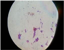
Figure 1 Microscopy of slides stained by Gram stain with presence of bacilli after 24 hours of incubation at 37ºC.
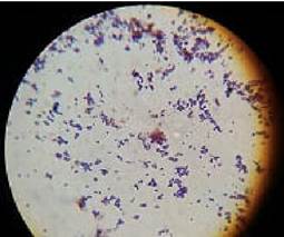
Figure 2 Microscopy of slides stained by Gram stain with presence of coconuts after 24 hours of incubation at 37ºC
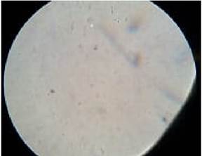
Of the antibiograms, after 24 hours of sowing of 10 samples in culture medium BHI and maintenance in gases the bacteriological 37ºC, were made slides by the method of Gram staining in order to research the growth of bacteria and identified a discrete presence of coccus in accordance with the characteristics morpho-dyeing, in tubes containing concentrations of 50 and 60% (Figure 3). However were not identified microorganisms at concentrations of 70, (Figure 4) 80 and 90%.
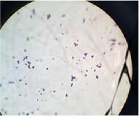
Figure 3 Microscopy of blade of the antibiogram with 60% where there was a slight presence of coccus.
Of the borderline 60% e 70% were prepared Petri plates containing Agar Müeller Hinton for each concentration for against proof and not identified growth of colonies in both concentrations (Table 1, Figure 5).
Table 1 Results of Petri plates used as evidence against
| Borderline | Results |
|---|---|
| 60% (Containing 1ml of extract of geopropolis) | Without growth |
| 70% (Containing 0.5 ml of extract of geopropolis) | Without growth |
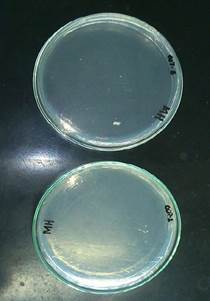
Figure 5 Petri plates with sowing of antibiogram of concentrations of 60% and 70% respectively, without growth of colonie.
This result suggests that there is antimicrobial activity of geopropolis of Melípona quadrifasciata at concentrations above 60%.
DISCUSSION
In this experiment were identified bacilli, Gram-positive and Gram-negative cocci, agreeing with Tilley, Smith Jr 1, Patel, Forsythe, Smith 2 e Oliveira et al 3.
After antibiograms, were not identified microorganisms at concentrations of 70, 80 and 90% different outcome than says Park, Ikegaki 16 about the concentration of 80% has the best results. In this experiment, there was inhibition in lower concentrations than that suggested by the authors.
The result obtained in this experiment suggests that the alcoholic extract of geopropolis of Melipona quadrifasciata has active components against some microorganisms often found in recurrent external otitis of dogs, similar to that encountered by Cunha 13, Campos et al 14 and Araújo et al 15, working with other species of this genus of stingless bees.
In conclusion, the geopropolis bee Melipona quadrifasciata showed efficacy in vitro against the microorganisms identified in this experiment and may be an alternative for the treatment of these recurrent otitis.
More tests are needed in order to find the best concentrations of geopropolis of Melipona quadrifasciata with antimicrobial activity.

