


 texto em
  Inglês (pdf)
 Artigo em XML
 Referências do artigo
 Enviar este artigo por email
 Citado por SciELO
  Citado por Google
  Similares em
SciELO
Similares em
SciELO  Similares em Google
 Permalink
Introduction
Autoimmune hepatitis (AIH) is a liver disease of unknown etiology that can affect people of all ages; its clinical presentation may be asymptomatic, chronic, or even debut as acute liver failure 1. Its prevalence in adults ranges from four to 42.9 per 100,000 people 2,3; with a 72-95% predominance in women 4,5.
Other autoimmune diseases have been found in 20-78% of patients with AIH 6,7, with autoimmune thyroiditis, ulcerative colitis and type 1 diabetes mellitus most commonly reported 6. Connective tissue diseases are less frequently associated with AIH, with rheumatoid arthritis and Sjogren's syndrome predominating in 1-4% of patients. Less frequently, systemic sclerosis (SS) has been associated with the disorder, with only 14 cases published in the literature 6.
Aware of the diagnostic difficulty related to the atypical presentation of AIH and the overlapping with other disorders 8, we present the case of a woman diagnosed with AIH and SS, her clinical findings, diagnosis and management.
Clinical case
This was a 42-year-old woman from a rural area in Santander, Colombia, with no significant medical history who presented with a three-month history of a growing, painless abdominal mass in the right upper quadrant, associated with self-limited weekly episodes of moderate rectal bleeding and anal itching (without predominance at any particular time of day), along with unquantified weight loss. She had consulted repeatedly at a lower care level where she received parasite treatment without resolving her symptoms.
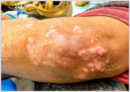
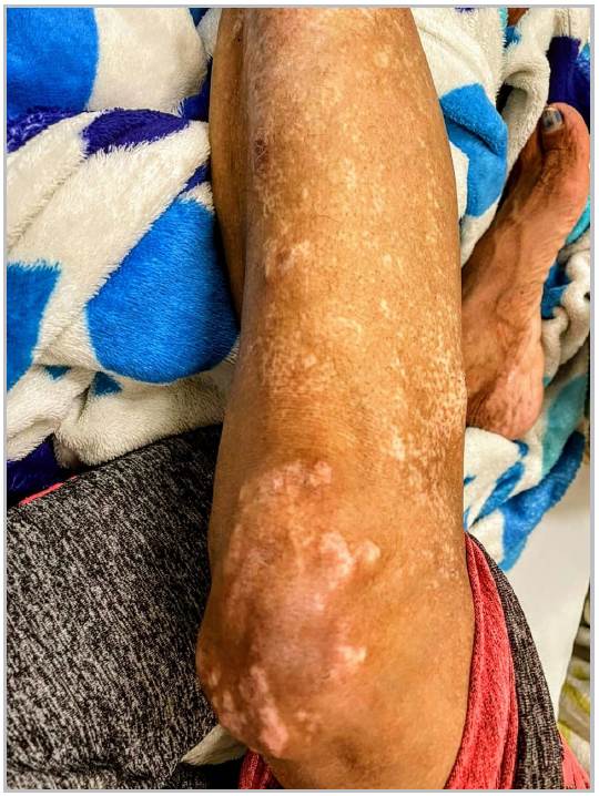
On physical exam, she was hemodynamically stable and hydrated, with no signs of systemic inflammation, a non-distended abdomen with no pain on palpation, a right upper quadrant 10 x 5 cm painless mass, and a liver edge 2 cm from the costal margin. There was no evidence of hemorrhoids on rectal palpation, with fecal matter in the rectal ampulla, and no lesions or bleeding palpated. Calcinosis cutis was found on the extensor surface of the elbow (Figure 1), along with salt-and-pepper depigmentation of the extremities (Figure 2), sclerodactyly and Raynaud's phenomenon (Figure 3).

On admission, the complete blood count revealed bicytopenia due to thrombocytopenia and leukopenia, and the ultrasound findings suggested chronic liver disease and moderate splenomegaly with indirect signs of portal hypertension. Therefore, in the context of a possible nonalcoholic cirrhosis, complementary tests were ordered, finding negative serology for hepatotropic viruses; a normal iron kinetics profile, albumin, total protein, C3, C4, bilirubin and coagulation times; elevated transaminases; positive IgM immunoglobulin, ANA and SMA; and negative anti-mitochondrial (AMAs) and anti-Scl-70 antibodies. A liver biopsy reported no fibrous expansion in the parenchyma with mild lymphocytic inflammatory infiltrate with occasional eosinophils mixed in. These findings were compatible with chronic hepatitis with mild necroinflammatory activity without fibrosis. She was therefore diagnosed with autoimmune hepatitis, which was also thought to overlap with limited cutaneous SS, and started on immunosuppression with azathioprine and prednisolone, with a satisfactory clinical response and decreased transaminase levels after four months of follow up.
Discussion
Autoimmune hepatitis is diagnosed using histopathological, clinical and paraclinical information, the latter including transaminitis, serum IgG elevation, and the presence of characteristic antibodies 1. However, as there is no diagnostic marker, finding AIH is a challenge, requiring a suspicion of the disease based on suggestive signs and symptoms and ruling out differential diagnoses, such as viral hepatitis, hereditary liver conditions and other autoimmune diseases including primary biliary cholangitis (PBC) and primary sclerosing cholangitis, with exceptions in the case of overlap syndromes 8.
Beginning in 1999, the diagnostic criteria endorsed by the International Autoimmune Hepatitis Group (IAIHG), have been proposed, with their latest simplified version, from 2008, indicating a score over seven as probable AIH, and liver biopsy being the fundamental element in the definitive diagnosis of the disease 9. In our case, three of the proposed criteria were met (antibodies, histopathology, lack of viral hepatitis), for a total of seven points, with a compatible histopathological report.
As for systemic sclerosis, it is recognized as a rare disease characterized by skin, subcutaneous tissue and internal organ fibrosis, as well as vascular disease 10. It is mainly classified clinically, with the fulfillment of the 2013 ACR/EULAR criteria 11, and diagnosis made beginning at a score of nine. After applying the classifying criteria, a score of 12 points was obtained (skin sclerosis on the fingers of both hands with proximal extension toward the metacarpophalangeal joints, sclerodactyly and Raynaud's phenomenon). Systemic sclerosis is classified as limited, diffuse and without scleroderma, based on the degree of skin involvement 11. In its limited type, SS can present clinically with the CREST syndrome, which is characterized by calcinosis, Raynaud's phenomenon, sclerdodactyly, esophageal dysmotility and telangiectasias, with the latter two not found in the reported case 12.
A low prevalence of liver involvement has been found in patients with SS; Abu-Shakra et al. found a prevalence of chronic liver disease of 1.5% in 262 patients 13. Likewise, cirrhosis findings have been documented in approximately 9% of autopsies on patients with SS 14.
Autoimmune hepatitis has been reported less frequently in patients with SS than PBC 15, which has been found in almost 10% of cases 16. In their retrospective study of 1,572 patients with SS, Marí-Alfonso et al. reported that 1.2% had AIH 17, with 14 cases of overlap of both diseases published so far 18-31 (Table 1). In these case reports, as well as our own, the presence of limited SS has been the most common (10/16 patients). The diagnosis of AIH has mainly occurred after finding SS, unlike the case of our patient and six other cases in which the diagnosis was concomitant. In almost all cases, the diagnosis was made after age 50, with one overlap case reported at age 17, associated with a diagnosis of polymyositis and sarcoidosis.
Although the pathophysiological relationship between AIH and SS is not clearly established, patients with AIH could share common autoimmune pathways with SS, as anticentromere antibodies are found in AIH cases 30. However, in light of the scant evidence to prove this, the need for more studies to confirm this hypothesis has been raised. Likewise, liver involvement could precede skin manifestations, and an evaluation for SS is recommended in patients with AIH 20,23.
The treatment of choice for patients with AIH is based on corticosteroids associated with azathioprine as a steroid sparing agent 32. In this case, as in all reported cases, liver profile normalization and modulation of the disease after adhering to steroid treatment were evident. None of the published cases had scleroderma renal crisis or risk in patients with SS receiving high-dose corticosteroids 33.
Regarding the prognosis of patients with SS, a 71.7-93% 10-year survival rate has been found in various cohort studies 34-36. Likewise, an 85-95% survival of AIH cases has been estimated for the same time frame 37,38, with poor prognostic factors under study including cirrhosis before or during treatment, severe liver dysfunction and treatment failure 39. There are no studies to date on the prognosis for patients with overlapping AIH and SS.
Table 1 Immunological characteristics of the published cases with SS and AIH.
| Author (Ref.) | Age | Sex | Autoimmune liver disease | Connective tissue disease | AMA | ACA | ANA | LKM | ASMA | IGG | Anti- dsDNA |
|---|---|---|---|---|---|---|---|---|---|---|---|
| Yabe et al. 18 | 51 | F | Concomitant AIH | LSS | - | + | + (1/640) | NT | - | + | - |
| Mamoru et al 19 | 48 | F | AIH five years after SS | LSS | - | + | + | NT | + | + | - |
| Marie et al. 20 | 67 | F | AIH seven years after SS | LSS | - | + | + (1/600) | - | - | + | NT |
| 48 | F | Concomitant AIH | LSS | - | + | + (1/1000) | - | - | + | NT | |
| Margaret et al. 21 | 47 | F | AIH-PBC. SS one year after AIH | LSS | NT | NT | + (1/640) | NT | + | NT | - |
| Lis-Swiety et al. 22 | 17 | F | AIH. SS one year after AIH | SS, polymyositis, sarcoidosis | NT | NT | + | NT | + | + | NT |
| Rodrigues et al.23 | 47 | F | AIH. After two years of SS | DSS | + | + | + | - | - | NT | NT |
| Efe et al. 24 | 60 | F | AIH-PBC. Six years after SS | LSS | + | + | + (1/320) | + | - | NT | NT |
| Klein et al. 25 | 47 | F | AIH. SS five years later | DSS | - | - | - | - | + | + | NT |
| You et al. 26 | 51 | F | Concomitant AIH | LSS | - | + | + (1/260) | - | - | + | NT |
| Pamfil et al. 27 | 53 | F | AIH. Three years after SS | LSS, polymyositis, cerebral vasculitis | - | + | + (1/1280) | - | - | + | - |
| Assandri et al. 28 | 70 | F | Concomitant AIH | DSS | - | NT | + (1/1280) | - | + | NT | NT |
| 55 | F | AIH. SS eight months later | LSS | NT | NT | + (1/1280) | NT | + | NT | NT | |
| Coelho et al. 29 | 40 | F | Concomitant AIH | DSS | NT | NT | - | NT | + | + | - |
| Toyoda et al. 30 | 58 | F | AIH-PBC. After SS | LSS and PTI | + | - | +(1/160) | NT | NT | + | + |
| Han et al. 31 | 41 | F | Concomitant AIH-PBC. | DSS | - | NT | + (1/640) | - | NT | + | NT |
| F: Female, M: Male, AIH: Autoimmune hepatitis, LSS: Limited systemic sclerosis, DSS: Diffuse systemic sclerosis, PBC: Primary biliary cholangitis, SS: Systemic sclerosis, AMA: Anti-mitochondrial antibodies, ACA: Anti-cardiolipin antibodies, ANA: Anti-nuclear antibodies, ASMA: Anti-smooth muscle antibodies, IGG: IgG antibodies, Anti dsDNA: Anti-double stranded DNA antibodies. | |||||||||||
Conclusions
Patients with AIH are at high risk for other autoimmune diseases which could worsen the disease course. Therefore, although it has not been reported much, the follow up, suspicion and evaluation of SS could lead to an early diagnosis and comprehensive treatment of cases with both conditions. Likewise, the clinical and paraclinical signs of liver involvement in patients with SS, with no known secondary cause, should trigger an investigation looking for an underlying autoimmune liver disorder.

