


 Inglês (pdf)
 Artigo em XML
 Referências do artigo
 Enviar este artigo por email
 Citado por SciELO
  Citado por Google
  Similares em
SciELO
Similares em
SciELO  Similares em Google
 Permalink
INTRODUCTION
Neospora caninum is an apicomplex protozoan, originally identified in the tissue of dogs with paralysis, and first identified in 1984 1-3, as an important cause of neurological disease in dogs worldwide 4. Neosporosis is one of the diseases that causes abortion in cows, sheep, goats, buffalo and camels; with special importance in bovines worldwide due to the important economic impact on milk and meat production 5.
N. caninum has a wide range of hosts. Natural infection has been evidenced in dogs, cows, sheep, goats, horses and deer. The experimental infection has been induced in mice, rats, dogs, foxes, cats 6, coyotes, pigs, gerbils, rabbits, sheep and cows 7.
Over time, N. caninum was confused with T. gondii. One of the reasons is that tachyzoites and cysts with bradyzoites, among both protozoa are very similar to light microscopy 8.
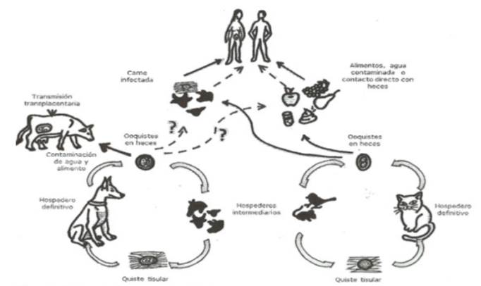
Several molecular studies have indicated a close phylogenetic relationship between N. caninum and T. gondii, both are obligated intracellular pathogens capable of infecting all types of nucleated cells in intermediate hosts, differentiating from each other by the final host being the cat for cases of T gondii and the dog for the case of N. caninum9,10, as shown in Figure 1.
The importance of T. gondii infection lies in its impact on public health as an opportunistic pathogen of immunocompromised individuals and in maternal-fetal infections, where children can be born with different neurological problems such as loss of vision or even death. On the other hand, despite its resemblance to T. gondii, N. caninum is not yet considered a pathogen that affects humans. Likewise, T gondii provides a study model for intracellular parasitism 11,12.
For the diagnosis of N. caninum there are several serological tests that can be used to detect antibodies against this protozoan, including ELISA, IFAT indirect immunofluorescence and direct agglutination tests. The presence of specific antibodies in the serum of cows with abortions indicates an exposure to N. caninum. For the final diagnosis, a diagnosis should be made in the aborted fetus using histopathology, immunohistochemistry and polymerase chain reaction (PCR) techniques. Currently there are different techniques to detect N. caninun DNA 13, using real-time PCR protocols (qPCR) 14.
Inoculation with N. caninum of nonhuman primates in gestation produces the transplacental transmission of the parasite and the induction of fetal encephalitis with lesions similar to those induced by transplacental infections of T. gondii in primates 15 There are reports on human exposure to N. caninum using IFAT, however, further trials are required to determine the significance of exposure and possible cross-reactions that may affect these results 16.
Currently, there are multiple diseases not etiologically recognized, that cause pathologies in humans due to the lack of diagnostic methods that unequivocally identify them. In the case of N. caninum, it is not yet known what effects on human health, it may have, which specific group affects, or which characteristic signs are likely to show up. Studies carried out in humans have focused on searching for similar signs of those known for T. gondii due to their biological closeness, such as abortion, symptoms in immunocompromised patients or in population with exposure factors to the agent (veterinarians, breeders). The signs may not be those considered a priori, but could be the same presented in their final host. Therefore, it should be studied under different circumstances and with other implications. Such as in humans with epidemiological risk factors conducting studies between exposed and not exposed to the infection.
It is necessary to have more clarity about the state of N. caninum in Colombia, and to carry out investigations that involve the animal species in its cycle with the possibility of adding human beings, considering the link between both species.
Therefore, the objective of this review is to analyze the zoonotic potential of Neospora caninum taking into account its biological relationship with T. gondii.
METHODOLOGY
A systematic review was carried out, using two databases for the collection of information (Science direct and SciELO), a search engine (Google Scholar) and the Repository of the Universidad Nacional de Colombia, all documents were organized by means of the bibliographic manager Mendeley for proper review. The search keywords used in Spanish in DeCS, BIREME were “Neosporosis”, “Neospora caninum”, “Toxoplasmosis”, “Toxoplasma gondii”, “Neospora humans” and in English in MeSH, NLM they were “Neospora human infection” “,” Neospora zoonotic “. This search, turned around 3,000 results for N. caninum and an average of 17,000 for T. gondii. Among the results, those related to diagnosis and studies in humans were selected. Forty documents were found that met these criteria and were divided into review articles, research and summaries. Of these, 24 were used for the introduction and discussion. Finally, 8 were selected for evaluating the infection in humans. The time period of the data collection was from 1998 to 2017.
RESULTS
8 documents, which are indicated in Table 1, met the inclusion requirements for the purpose of this study (studies that analyzed the infection in human samples). In these works, tests were performed to look for antibodies against N. caninum in humans, but none used methods to isolate or cultivate the parasite.
Table 1 Studies for the detection of anti-N. caninum antibodies in sera and cell lines in humans.
| Authors | Method | Number human samples | % positivity |
|---|---|---|---|
| Ho-Woo et al (17) 1998 | ELISA, Western blot and IFAT. | 172 sera (+) T. gondii 110 sera (-) T.gondii | 6.7% cross reaction, 3/172 (1.7%) and 1/110 (0.9%) for N, caninum |
| Petersen et al (18) 1999 | ELISA, Western blot and IFAT. | 76 sera of women with repeated miscarriages | 1.3% |
| Tranas et al (19) 1999 | IFAT and Western blot. | 1029 serum samples | 6.7% 1:100 |
| Lobato et al (20) 2006 | IFAT, ELISA, Western blot. | 256 serum samples: 61 VIH (+), 50 patients with neurological signs, 91 newborns, 54 healthy | 38%, 18%, 5% and 6%, respectively |
| McCann et al (21) 2008 | ELISA, IFAT. | 3232 samples of general population serum and 518 high risk group | 21.38% and 5.56% >20% ELISA inhibition |
| Gangneux et al (22) 2009 | IFAT and ELISA | 500 sera in healthy women and 400 VIH (+)sera | 8% healthy, 4% VIH (+) => 1:20; 0.7%=> 1:80; 0.2%=> 1:160 |
| Benetti et al (23) 2010 | IFAT, Western blot | 1036 samples of serum : 932 bovine dairy females, 37 dogs 67 humans | (53.5%), (67.6%) (10.5%), respectively, |
| Carvalho et al (24) 2010 | LDH and MTT, ELISA | Cell lines BeWo and HeLa | Susceptibility of infection in both cell lines |
Ho-Woo 17, detected anti-N. caninum antibodies in positive and negative human sera against Toxoplasma gondii by ELISA, Western blot and IFAT. Twelve sera of 172 (6.7%) positive for T. gondii cross-reacted with N. caninum antigens, and one of 110 negative sera for T. gondii (0.9%) reacted with N. caninum antigens by ELISA. By western blot, the 12 sera reacted against T. gondii with several band patterns 30kDa (SAG1) and 22 kDa (SAG2), but with N. caninum the number of reactive bands decreased. At 43 kDa (SAG), three cases of the T. gondii positive group and one of the T. gondii negative group reacted. By IFAT all sera from the T. gondii positive group marked the membrane surface for T. gondii, whereas for N. caninum the fluorescence was detected at the membrane surface, cellular organelles, or both. The group of T. gondii negative also reacted strongly for N. caninum in cellular organelles; suggesting that antibodies against N. caninum may be present in human sera, however, the rate of positives to infection was very low 3/172 (1.7%) and 1/110 (0.9%).
Petersen 18, determined whether N. caninum, known to cause repeated miscarriages and stillbirths in cattle, also causes repeated abortions in women. To do so, they retrospectively examined serum samples from 76 women with an average age of 30.8 years, with a history of repeated miscarriages and intrauterine fetal death for probable evidence of N. caninum infection. In this study, no antibodies against the parasite were detected by ELISA, IFAT or western blot.
Tranas 19 searched for evidence of human exposure to N. caninum by detecting antibodies in blood donors by IFAT and western blot. Of the 1,029 samples examined, 69 (6.7%) had 1: 100 titles through IFA tests. Fifty of the 69 (72%) sera that were positive for N. caninum were negative for T. gondii. Immunoblot analysis confirmed the specificity of sera positive for N. caninum antigens with several sera recognizing multiple N. caninum antigens with molecular masses similar to those of antigens recognized for serum with antigens against N. caninum in non-human primates. An immunodominant antigen of approximately 35 kDa was observed in 12 sera. These data provide evidence of human exposure to N. caninum although antibody titers in healthy donors were low, ignoring the significance of human exposure and possible infection with this parasite.
Lobato 20, investigated the presence of N. caninum antibodies in seronegative individuals and seropositive to T. gondii by means of IFAT, ELISA and Western blot. A total of 256 serum samples divided into four groups were evaluated: 61 samples of HIV-positive patients, 50 samples of patients with neurological disorders, 91 samples of newborns and 54 samples of individuals. Immunoglobulin G N. caninum antibodies were predominantly detected in HIV-infected patients (38%) and patients with neurological disorders (18%), while newborns and healthy individuals showed lower percentages of seropositivity (5 and 6). %, respectively). Seropositivity to N. caninum was significantly associated with seropositivity to T. gondii both in patients infected with HIV and in patients with neurological disorders. Seroreactivity to N. caninum was confirmed by Western blot, with positive sera predominantly recognizing the 29 kDa antigen of N. caninum. The results of this study indicate the presence of infection or exposure of N. caninum in humans, particularly in HIV-infected patients or patients with neurological disorders, who could have opportunistic and concurrent infections with T. gondii.
McCann 21, in England, performed retrospective tests of 3232 serum samples from the general population and 518 serum samples from a high-risk group using IFAT and ELISA to analyze the frequency distribution. There was no evidence of human exposure to N. caninum.
Gangneux 22 investigated the seroprevalence of N. caninum in humans in France, where the seroprevalence for T. gondii is high. They used 500 sera from healthy women and 400 serum samples from patients infected with HIV. All serum samples were subjected to antibody tests against T. gondii by IFAT and ELISA. In 40 samples (8%) of immunocompetent persons and in 21 samples (4%) of immunocompromised persons, a weak fluorescence was observed at a 1:20 dilution. None of the 500 samples from immunocompetent patients was positive for anti-caninum antibodies when evaluated at a dilution of 1:80. Within the group of immunocompromised people, 3 were seropositive for N. caninum at a titer of 1: 80 and one sample was positive with a titer of 1: 160. No evidence of infection or exposure of N. caninum was found in immunocompetent persons, but possible infection by N. caninum could not be excluded.
Benetti 23, evaluated the frequency of antibodies against N. caninum in dairy cattle of the southwestern region of the state of Mato Grosso, Brazil, in addition to serum samples obtained from dogs and humans living on farms. 1036 serum samples were analyzed, of which 932 (89.9%) were from dairy bovine females, 37 (3.57%) from dogs and 67 (6.46%) from humans, who lived in 24 farms and were examined by IFAT. The reactive samples of human serum were again analyzed by Western blot to confirm the results. Antibodies against N. caninum were found in 499 cattle sera (53.5%), with at least one positive in each farm, 25 dog sera (67.6%) and seven human sera (10.5%). There were no significant differences in the number of positive bovine sera according to age group.
Carvalho 24, verified the susceptibility of human trophoblastic cells (BeWo) in comparison with cervical uterine cell lines (HeLa) before infection with N. caninum. BeWo and HeLa cells were infected with different concentrations of the parasite: N. caninum tachyzoites and analyzed at different times after infection to observe cell viability. Both cell lines were also evaluated for cytokine production and parasite infection / replication assays when pre-treated or not with N.caninum lysate antigen (NLA) or recombinant human IFN-γ. Cell viability increased up to 48 h post-infection in both cell types, suggesting that the infection could inhibit early cell death and / or induce cell proliferation. The HeLa cells were more susceptible to infection by N. caninum than the BeWo cells and the previous treatment with IFN-γ showed a reduced infection index in both cell lines. It was concluded that BeWo and HeLa cells were infected by N. caninum, although they show differences in susceptibility to infection, cytokine production and cell viability.
DISCUSSION
Zoonoses continue to register high incidence rates in countries causing significant morbidity and mortality. Cattle infections and parasitosis alter organic functioning and reduce their productive index. Humans become infected through food, water, soil and close contact with animals. These diseases are also an obstacle to international trade, as well as a serious economic deficit for livestock industries and, in general, for the economy of a community or country, which can have wide repercussions for public health in a society 25. Highlighting the very close morphological and biological phylogenetic relationship that exists between T. gondii and N. caninum9, and that in the beginning, cases in animals such as cattle and canines infected with N. caninum clinically and pathologically could be misdiagnosed as infected by T. gondii (19), the possibility of N. caninum infection in humans should be considered, given its biological similarity with T. gondii and considering its clinical importance. Three studies so far show exposure of N. caninum in humans; serologically positive samples by means of ELISA, IFAT and western blot in patients with HIV, with neurological signs or with predisposing factors such as a direct link with bovine production and contact with dogs in rural areas 19,20,23.
Research should continue to search for N. caninum infection in humans by serological tests or alternatively, using tissue obtained from biopsy to be processed by PCR or immunohistochemistry 17.
Large-scale studies involving different countries are needed to more accurately determine the potential role of this parasite in immunocompetent humans with defined and immunodeficient risk factors to isolate the parasite or detect N. caninum DNA in these patients. It is also convenient to carry out more studies to identify the surface proteins of N. caninum and thus avoid possible cross-reactions with surface proteins of T. gondii.
The lack of evidence of N. caninum infection in women who had repeated spontaneous abortions does not rule out the possibility that the infection could occur in humans. The predominant effects of neosporosis in dogs are mainly progressive neurological signs that include paralysis. Therefore, it may be useful to examine human patients with clinical symptoms other than abortions, for example, neurological disorders of unknown etiology 18. In this regard, the human being could be exposed to N. caninum as an infectious form or simply an antigenic form.
Several in vitro studies where human cells have been used as host cells, such as fibroblasts 26, human foreskin 27 human colon tumor cells 28 and human mammary gland 29, may be evidence of infection with N. caninum. These results support the notion that N. caninum can invade a variety of cell types from different hosts, including cattle, dogs, cats, mice, sheep, monkeys and human cells.
With the evidence obtained so far, the role of N. caninum as a human pathogen is unclear. Serological data from humans only suggest that infection could occur. Studies would be required, for example, with molecular techniques that allow the species to be sequenced and confirmed, since immunological techniques will always have the possibility of cross-reaction. 30
It is necessary to carry out more epidemiological studies of the disease, obtaining seroprevalences in different farms, evaluating all the predisposing factors altogether, obtaining samples of cattle, dogs and humans as in the study done by Benedetti 23 and thus getting a general perspective of the disease in the country and worldwide. Considering that dogs are both the intermediate host and the definitive host of N. caninum, these could represent a potential threat for humans due to their close relationship 31,32.
It is necessary to carry out more epidemiological studies, obtaining seroprevalence figures at different regions and to evaluating all the possible risk factors involved, as well as obtaining samples from humans, bovine and dogs as performed previously by Benedetti 23. In this way it would be obtained a general perspective of the situation of this infection in Colombia and the rest of the world. It should be taken into account that dogs are the definitive host and because its near relation with humans, represent a potential zoonotic risk 31,32.
N. caninum is an intracellular protozoan closely related to T. gondii, as demonstrated by the comparison of the genomes of both organisms 11. It is not ruled out that the probability of zoonosis due to N. caninum infection may exist, but more research is required to demonstrate the level of exposure, risk factors and associated clinical signs or pathologies.

