SciELO - Scientific Electronic Library Online

 
vol.36 issue4Effect of weaning weight on growth performance, feed efficiency, and behavioral characteristics of Holstein-Friesian and Brown Swiss calvesEffect of dietary nano α-tocopherol acetate plus selenium on productivity, immune status, intestinal morphometry, and carcass traits of broiler chickens author indexsubject indexarticles search
Home Pagealphabetic serial listing  

Services on Demand

Journal

Article

Indicators

Related links

  • On index processCited by Google
  • Have no similar articlesSimilars in SciELO
  • On index processSimilars in Google

Share


Revista Colombiana de Ciencias Pecuarias

Print version ISSN 0120-0690On-line version ISSN 2256-2958

Rev Colom Cienc Pecua vol.36 no.4 Medellín Oct./Dec. 2023  Epub Aug 14, 2024

https://doi.org/10.17533/udea.rccp.v36n4a2 

Accepted Manuscripts

Oxidative stress parameters in dogs naturally infected with sarcoptic mange

Parâmetros de estresse oxidativo em cães naturalmente infectados com sarna sarcóptica

Parámetros de estrés oxidativo en perros naturalmente infectados con sarna sarcóptica

Kerim Emre Yanar1  * 

Sefa Kucukler2 

Emre Eren3 

Muhammed Sertaç Eroglu4 

Murat Ilgun5 

Cihan Gur6 

Fatih Mehmet Kandemir7 

Mustafa Sinan Aktas8 

1Ataturk University, Department of Internal Medicine, Faculty of Veterinary Medicine, Erzurum, Turkey, emre.yanar@atauni.edu.tr, https://orcid.org/0000-0001-7302-7077

2Ataturk University, Department of Biochemistry, Faculty of Veterinary Medicine, Erzurum, Turkey, https://orcid.org/0000-0002-8222-5515

3Ataturk University, Department of Internal Medicine, Faculty of Veterinary Medicine, Erzurum, Turkey, https://orcid.org/0000-0002-2966-5466

4Ataturk University, Department of Internal Medicine, Faculty of Veterinary Medicine, Erzurum, Turkey, https://orcid.org/0000-0003-4243-8273

5Ataturk University, Department of Internal Medicine, Faculty of Veterinary Medicine, Erzurum, Turkey, https://orcid.org/0000-0002-0062-2825

6Ataturk University, Department of Biochemistry, Faculty of Veterinary Medicine, Erzurum, Turkey, https://orcid.org/0000-0001-6775-7858

7Aksaray University, Department of Biochemistry, Faculty of Medicine, Aksaray, Turkey, https://orcid.org/0000-0002-8490-2479

8Ataturk University, Department of Internal Medicine, Faculty of Veterinary Medicine, Erzurum, Turkey, https://orcid.org/0000-0002-7206-5757


Abstract

Background:

Scabies is one of the most common diseases in dogs. It threatens both animals and humans due to its zoonotic potential.

Objective:

The purpose of this study was to evaluate the oxidant/antioxidant balance with hematological findings in dogs infested with sarcoptic mange.

Methods:

The animals evaluated in this study consisted of 32 mixed-breed dogs between 1 and 2 years of age. The dogs were allocated into two groups: a control group (infestation-free animals; n=10), and a sarcoptic mange-infected group (Sarcoptes, n=22). Dogs in the Sarcoptes group showed infestation signs such as intense itching, excoriations, alopecia, and blistering of the elbow and auricular margins.

Results:

Significant increase (p<0.01) levels were observed in total oxidant status (TOS), malondialdehyde (MDA), oxidative stress index (OSI), and nitric oxide (NO), while glutathione (GSH) and total antioxidant status (TAS) levels in dogs infested with Sarcoptes decreased significantly (p<0.01). In addition, a significant increase (p<0.01) of WBC count in dogs in the sarcoptic group in comparison with the control was found. Conversely, there was significant decrease (p<0.01) in RBC, HGB, and PCV counts in Sarcoptes-infested dogs.

Conclusions:

Our study suggests a possible relationship between oxidant/antioxidant imbalance and hematological findings in dogs infested with sarcoptic mange. Furthermore, in addition to MDA, TAS, TOS, and OSI markers, NO as well as GSH might be also used to assess the oxidative stress in dogs naturally infected with Sarcoptes scabiei.

Keywords: biomarker; dog; infestation; oxidative stress; Sarcoptes; sarcoptic mange; scabies; zoonosis

Resumen

Antecedentes:

La sarna es una de las enfermedades más comunes en perros. Es una amenaza para la salud animal y humana por su carácter zoonótico.

Objetivo:

El propósito de esta investigación fue estudiar el balance oxidante/antioxidante con hallazgos hematológicos en perros infestados por sarna sarcóptica.

Métodos:

Los perros del grupo Sarcoptes presentaban signos de infestación tales como prurito intenso, excoriaciones, alopecia y ampollas en el codo y márgenes auriculares. El estudio evaluó 32 perros mestizos entre 1 y 2 años de edad. Los perros se distribuyeron en dos grupos: un grupo control (animales libres de infestación; n=10), y un grupo con sarna sarcóptica (Sarcoptes; n=22).

Resultados:

Se encontró un aumento significativo (p<0.01) en el estado oxidante total (TOS), malondialdehído (MDA), índice de estrés oxidativo (OSI) y niveles de óxido nítrico (NO), mientras que el glutatión (GSH) y los niveles de estado antioxidante total (TAS) de los perros infestados por Sarcoptes disminuyeron significativamente (p<0.01). Además, se encontró un aumento significativo (p<0,01) en el recuento de leucocitos de los perros del grupo Sarcoptes en comparación con el control. Por el contrario, hubo disminuciones significativas (p<0,01) en los recuentos de RBC, HGB y PCV de los perros infestados.

Conclusiones:

Podría sugerirse una posible relación entre el desequilibrio oxidante/antioxidante y los hallazgos hematológicos en perros infestados por sarna sarcóptica. Adicionalmente, los marcadores MDA, TAS, TOS y OSI, el NO y el GSH podrían utilizarse para evaluar el estrés oxidativo en perros infectados naturalmente por Sarcoptes scabiei.

Palabras clave: biomarcador; escabiosis; estrés oxidativo; infestación; perro; Sarcoptes; sarna sarcóptica; zoonosis.

Resumo

Antecedentes:

A sarna é uma das doenças mais comuns em cães, e também ameaça a saúde humana e animal devido ao seu caráter zoonótico.

Objetivo:

O objetivo da pesquisa foi estudar o equilíbrio oxidante/antioxidante com achados hematológicos em cães infestados com sarna sarcóptica.

Métodos:

Os cães do grupo Sarcoptes apresentavam sinais de infestação como prurido intenso, escoriações, alopecia e bolhas no cotovelo e margens auriculares. O material deste estudo consistiu em 32 cães sem raça definida, entre 1 e 2 anos de idade. Os cães foram divididos em dois grupos que foram grupo controle (animais livres de infestação, n=10), bem como o grupo sarna sarcóptica (Sarcoptes; n=22).

Resultados:

Os resultados do estudo demonstraram que houve aumentos significativos (p<0,01) no total oxidant status (TOS), malondialdeído (MDA), oxidative stress index (OSI) e nos níveis de óxido nítrico (NO), enquanto glutationa (GSH) e os níveis de status antioxidante total (TAS) dos cães infestados por Sarcoptes diminuíram significativamente (p<0,01). Além disso, foi encontrado um aumento significativo (p<0,01) da contagem de leucócitos dos cães do grupo Sarcoptes em comparação com o controle. Por outro lado, houve reduções significativas (p <0,01) nas contagens de RBC, HGB e PCV em cães infestados por Sarcoptes. Conclusões: Pode-se sugerir uma possível associação entre desequilíbrio oxidante/antioxidante e achados hematológicos nos cães infestados por sarna sarcóptica. Além disso, além dos marcadores MDA, TAS, TOS e OSI, NO e GSH também podem ser usados para descobrir o estresse oxidativo em cães naturalmente infectados por Sarcoptes scabiei.

Palavras-chave: biomarcador; cachorro; estresse oxidativo; infestação; Sarcoptes; sarna sarcóptica; sarna; zoonose

Introduction

Scabies is an important problem threatening health of humans and animals (Mcclain et al., 2009). It was first detected microscopically in 1687 by the Italian physician Giovan Cosimo Bonomo. Linnaeus identified two different agents of scabies, one in animals and the other in humans (Ljunggren et al., 2005). Sarcoptes scabiei var canis, Otodectes spp., trombiculid mites, Cheyletiella yasguri as well as Demodex canis, are the causative agents of mange in dogs in tropical and subtropical regions (Hampel et al., 2018). Among all these, the agents S. scabiei and D. canis have significant roles in the deterioration of health and welfare of infected animals (Paterson et al., 2014). Sarcoptes scabiei, with a worldwide distribution, affects especially young domestic dogs. Sarcoptic mange causes severe skin lesions that can affect dog skin, mostly in the form of alopecia, thickened skin structure, intense itching, dry, exudative crusts, and hemorrhagic and non-hemorrhagic cracks on the facial skin, upper neck, and upper eyelids (Uzuegbu et al., 2015).

Pro-inflammatory cytokines such as Interleukin 1α, Interleukin 1β, Interferon-γ and Tumor necrosis factor-α have a remarkable role in the occurrence of the disease (Arlian et al., 1996; Lalli et al., 2004). Triggering of these cytokines leads to overproduction of reactive oxidants and free radicals including free reactive oxygen species (ROS) such as hydroperoxide radicals (OH), superoxide anion radicals (O2), and reactive nitrogen species such as nitric oxide (NO) (Bickers and Athar, 2006). Antioxidants have an important role in preventing cell damage by means of reducing free radicals (Kleczkowski et al., 2003). Healthy animals keep a balance between the antioxidant system and free oxygen radicals, and the change of the balance is known as oxidative stress (Ercan and Fidanci, 2012). In addition, when excessive ROS accumulation occurs along with insufficient antioxidant mechanism to neutralize free radicals the cellular function may change and biological damage may occur (Knight et al., 2000). Free radicals also cause adverse effects such as inflammation, edema, wrinkles, erythema, autoimmune reaction, keratinization abnormalities, and hypersensitivity on the skin. The tunneling activity in the skin caused by parasites in scabies is an important cause of oxidative stress (Camkerten et al., 2009).

Few studies have investigated oxidative stress in sarcoptic mange (De and Dey, 2010; Allaam et al., 2014; Beigh et al., 2016). Additionally, no study has evaluated malondialdehyde (MDA), total antioxidant status (TAS), oxidative stress index (OSI), glutathione (GSH), NO, and total oxidant status (TOS) markers in dogs with sarcoptic mange as well as RBC, HGB and PCV counts. Consequently, the evaluation of oxidative stress by using MDA, TAS, TOS, GSH, NO, and OSI markers along with some hematological parameters in dogs naturally infected with S. scabiei was aimed in this study.

Materials and Methods

A total of 32 mixed-breed dogs between 1 and 2 years of age (Ethics Committee Decision No: 185/2021) were evaluated in the study. The dogs were allocated into 2 groups which were control group (infestation-free animals; n=10), and a sarcoptic mange group (Sarcoptes; n=22). The dogs in the Sarcoptes group were naturally infected with sarcoptic mange and they showed infestation signs such as intense itching, excoriations, alopecia, and blistering of the elbow and auricular margins.

Scrapings (scraping area varying from 1to5 cm) from the center and peripheries of the lesions on different parts of the face, ear, neck, elbow, shoulder, and tail of dogs diagnosed with clinical scabies was taken into sterile containers. The samples were mixed with 5 mL 10% KOH to be free of tissue materials, and then two drops were taken and put on a slide to examine under a microscope at 10X magnification (Shalaby et al., 2016).

Blood samples were taken from vena cephalica antebrachium into 10 mL serum tubes (Vacutainer tube with clot activator, Becton Dickinson Co. USA) and sterile test tubes containing 0.14% anticoagulant (EDTA K3, Pty Ltd., Adelaide, SA, Australia). The samples were kept at room temperature during 30 min. Then they were centrifuged at 3,000 rpm×10 min, and sera samples were stored at -80 °C until analyses. For complete blood cell count, an Abacus® Junior Vet5 (Diatron In. Budapest, Hungary) automated blood cell counter was used (WBC, RBC, HGB, and PCV).

Malondialdehyde levels of sera samples were determined spectrophotometrically by the method of Placer and colleagues with modification (Placer et al., 1966). The thiobarbituric acid (TBA) assay was used to measure MDA levels in sera samples. A pink color was observed in TBA and MDA reactions and then the color was analyzed at 532 nm in the spectrophotometer. The MDA levels are expressed as nmol/g blood.

The Sedlak and Lindsay method was used to measure GSH levels of blood. For this purpose, 5,5’-Dithiobis (2-nitrobenzoic) acid is reduced in this method by compounds containing sulphydryl groups and the acquired yellow color was assayed at 412 nm in the spectrophotometer. The GSH levels are expressed as nmol/g blood (Sedlak and Lindsay, 1968).

A novel automated method developed by Real Assay Diagnostic (Turkey) was used to determine sera TAS levels. The oxidative reactions initiated by hydroxyl radicals in the mixture are inhibited by the antioxidant components in the sample, and inhibition of the color change and TAS are measured in the sera sample (Erel, 2004).

The TOS levels in sera were determined by using a novel automated method (Erel, 2005). The ferrous ion-O-dianisidine complex is converted to ferric ion by oxidants in the plasma sample. The oxidation reaction is enhanced by glycerol molecules plentifully available in the reaction medium. The ferric ion in an acidic medium forms a colored complex with xylenol orange. Color intensity is measured by spectrophotometry.

The OSI value was obtained with the following formula:

OSI = TOS (μmol H2O2 equivalent/L)/TAS (μmol Trolox equivalent/L) x100.

Sera NO levels were determined with a commercial test kit (Enzo Life Science, USA). The sera NO measurements are based upon the enzymatic conversion of nitrate to nitrite and the colorimetric detection of nitrite, a colored azo dye compound of the Griess reaction.

The SPSS statistics program (Statistical Package for Social science) was used for statistical analysis. Since preliminary results of Kolmogrov-Smirnov test revealed that data had normal distribution, a t-independent test was used for statistical comparisons of the treatment groups under study (SPSS, 2020).

Results

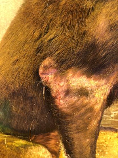
Clinical observations revealed lesions of erythematous skin with irregular alopecic areas along with scab formation. Skin lesions also had crusting and dermatitis, which appeared in various body areas of the Sarcoptes group (Figure 1). In addition, all dogs in the sarcoptic mange group had intense pruritus. We also observed that dogs in the control group had good appetite, body condition, as well as normal vital signs at clinical examination.

Figure 1 Crusting and dermatitis in the Sarcoptes group. 

Microscopic examination revealed similar alterations in all diseased dogs. However, their sizes changed depending on the intensity of the Sarcoptes infection (Figure 2).
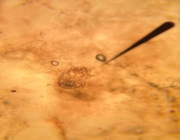
Figure 2 Sarcoptes scabiei var canis isolated from sampled dogs (10% potassium hydroxide preparation, X10 magnification). 

Mean values of MDA, GSH, TAS, TOS, OSI, and NO in control and infected groups are presented in Table I. In comparison with the control group, there were significant increase in MDA, NO, OSI, and TOS levels along with significant (p<0.01) decrease in GSH and TAS levels in the Sarcoptes infested dogs.

Table 1 MDA, GSH, TAS, TOS, OSI, and NO levels in healthy and infested dogs with sarcoptic mange. 

Biochemical parameters Groups N Mean Standard error of the mean Significance
MDA Control 10 2.0510 0.12084 **
Sarcoptes 22 3.9268 0.25065
GSH Control 10 0.4080 0.00998 **
Sarcoptes 22 0.2214 0.00703
TAS Control 10 1.8810 0.04418 **
Sarcoptes 22 1.3145 0.02569
TOS Control 10 4.4320 0.22611 **
Sarcoptes 22 6.6536 0.24930
OSI Control 10 2.3500 0.08539 **
Sarcoptes 22 5.0773 0.17978
NO Control 10 387.3000 7.28674 **
22 464.8759 8.66339

MDA: Malondialdehyde; GSH: Glutathione; TAS: Total antioxidant status; TOS: Total oxidant status; OSI: Oxidative stress index; NO: Nitric oxide; **: p<0.01.

Hematological findings in all dogs are presented in Table 2. Significant increase (p<0.01) in WBC count in dogs of the Sarcoptes group compared to the control group was observed. On the other hand, significant decrease (p<0.01) in RBC, HGB, and PCV counts in Sarcoptes-infested dogs were found compared with the control group.

Table 2 Hematological findings in control and dogs infested with sarcoptic mange. 

Hematological parameters Groups N Mean Standard error of the mean Significance
WBC Control 10 10.253 0.58423 **
Sarcoptes 22 13.419 0.75181
RBC Control 10 6.6 0.23224 **
Sarcoptes 22 5.5225 0.16845
HGB Control 10 14.91 0.57144 **
Sarcoptes 22 11.945 0.34607
PCV Control 10 44.514 1.43278 **
Sarcoptes 22 32.8305 .95314

WBC: White blood cell; RBC: Red blood cell; HGB: Hemoglobin; PCV: Pocket cell volume; **: p<0.01.

Discussion

Sarcoptic mange is the contagious skin disease in dogs caused by S. scabiei and is characterized by intense pruritus, alopecia, vesiculo-papular eruption, pinpoints crusts, and may cause death if left untreated (Kemp et al., 2002). In the present study, erythematous skin with irregular alopecia areas with crusting, and crusting and dermatitis on various body parts was observed. In addition, severe itching was present in all dogs in the sarcoptic mange group. The tunneling activity in the skin caused by parasites in scabies is seen as an important cause of oxidative stress (Mark et al., 2015). This study evaluated oxidative stress and changes in hematological parameters in dogs naturally infested with S. scabiei.

Free radicals are uninterruptedly created in the organism during normal metabolism. However, the production rates of free radicals increase even more in certain inflammatory disorders or syndromes. It is known that oxidative stress plays a significant role in the etiopathogenesis of various parasitic diseases in humans and animals (Chandramathi et al., 2009). Increased lipid peroxide is responsible for the pathology of skin lesions caused by Sarcoptes mites. Lipid peroxidation is an important cellular damage mechanism and is used as an indicator of oxidative stress in cells and tissues. Lipid hydroperoxides are by-products of lipid peroxidation and increased levels of lipid peroxidation products are closely associated with parasitic invasion (Kaya et al., 2007; CAM et al., 2008).

Malondialdehyde is the final compound of lipid peroxidation and is considered a key marker of oxidative stress. Increased MDA levels in peripheral blood showed exhaustion of enzymes and interrupted oxidative damage to the tissues of dogs with clinical sarcoptic mange. Parallel observations in mange cases were also described in other animal species, such as goats (De and Dey, 2010), pigs (Dimri et al., 2014), and camels (Espinosa et al., 2017).

Remarkably decreased levels of GSH, TAS, increased TOS and OSI level may be the result of overproduction of free radicals by the inflammatory cells recruited to combat the parasites, finally disrupting the antioxidant system of infested dogs. Similarly, Kocyigit et al. (2005) showed that intra and extra cellular parasites may induce or activate several oxidant-generating enzymes. Finally, inflammatory cell activation can occur in the organism. The increase in oxidant and decrease in antioxidants cannot be prevented in various diseases, and it has been reported that the oxidative/antioxidative balance shifts towards the oxidative state (Erel, 2005; Kocyigit et al., 2005; Cemek et al., 2006).

Increased NO production was also reported in humans with cutaneous leishmaniasis and other inflammatory skin diseases (Bickers and Athar, 2006). Free radicals are produced continuously by normal metabolic processes, but production rate increases during certain inflammatory or other disease conditions (Dimri et al., 2008). Once parasites are phagocytosed by macrophages, these cells produce ROS, such as O2− , H2O2, OH, and NO as a host defense mechanism (Serarslan et al., 2005). The NO increase may have been due to this mechanism.

As shown in Table 2, there were significant changes in various hematological parameters in the dogs affected by Sarcoptes mange. The dramatic decrease in circulating erythrocyte counts as well as hemoglobin amounts suggests an anemic condition. This result is quite like findings in camels (Parmar et al., 2005), goats (De and Dey, 2010), and sheep with mange (Aatish et al., 2007). It has been reported that anemia due to loss of skin proteins and leukocytosis might be due to an allergic reaction caused by mites or inflammatory reaction products (Pérez et al., 2015). The cause of anemia in the Sarcoptes group may be explained by loss of skin proteins and leukocytosis, as Pérez et al. (2015) stated.

The presence of S. scabiei on the host can cause itching and abrasions of the skin. This erosion can lead to invasion of bacteria and harmful microorganisms often causing an immune response characterized by leukocytosis (Stevanović et al., 2020). This seems to be the best explanation for the leukocytosis seen in this study.

Conclusion

Leukocytosis, anemia, and oxidative stress can occur in dogs naturally infected with S. Scabiei while MDA, GSH, TAS, TOS, OSI, and NO parameters can be used as diagnostic tools. However, further studies are needed to better explain the pathogenesis of sarcoptic mange infestation in dogs.

References

Aatish HU, Zia-Ud-Din S, Iqbal Z, Jabbar A, Tasawar Z. Prevalence of sheep mange in district Dera Ghazi Khan (Pakistan) and associated hematological/biochemical disturbances. IJAB 2007; 9(6):917-920. [ Links ]

Allaam MA, Allam TS, Elkhatam AO. Biochemical and circulating oxidative stress biomarkers in Egyptian buffaloes (Bubalus bubalis) infested by sarcoptic mange. Glob Vet 2014; 13(4):656-661. [ Links ]

Beigh SA, Soodan JS, Bhat AM. Sarcoptic mange in dogs: Its effect on liver, oxidative stress, trace minerals and vitamins. Vet Parasitol 2016; 227:30-34. https://doi.org/10.1016/j.vetpar.2016.07.013Links ]

Bickers DR, Athar M. Oxidative stress in the pathogenesis of skin disease. J Invest Dermatol 2006; 126(12):2565-2575. https://doi.org/10.1038/sj.jid.5700340Links ]

Cam Y, Atasever A, Erslan G, Kibar M, Atalay O, Beyaz L, Incı A, Lıman BC. Eimeria steidae: experimental infection in rabbits and the effect of treatment with toltrazuril and ivermectin. Exp Parasitol 2008; 119(1):164-172. https://doi.org/10.1016/j.exppara.2008.01.005Links ]

Camkerten I, Sahin T, Borazan G, Gokcen A, Das A. Evaluation of blood oxidant/antioxidant balance in dogs with sarcoptic mange. Vet Parasitol 2009; 161(1-2):106-109. https://doi.org/10.1016/j.vetpar.2008.12.019Links ]

Cemek M, Enginar H, Karaca T, Unak P. In vivo radio-protective effects of Nigella satival oil and reduced glutathi-one against irradiation-induced oxidative injury and number of peripheral blood lymphocytes in rats. Photochem Photobiol 2006; 82(6):1691-1696. https://doi.org/10.1111/j.1751-1097.2006.tb09832.xLinks ]

Chandramathi S, Kumar S, Sinnadurai S, Kuppusamy UR. Stress exacerbates infectivity and pathogenicity of blastocystis hominis. In vitro and in vivo evidence. Plos One 2014; 9(5):e94567. https://doi.org/10.1371/journal.pone.0094567Links ]

De UK, Dey S. Evaluation of organ function and oxidant/antioxidant status in goats with sarcoptic mange. Trop Anim Health Prod 2010; 42(8):1663-1668. https://doi.org/10.1007/s11250-010-9618-yLinks ]

Dimri U, Sharma MC, Swarup D, Ranjan R, Kataria M. Alterations in hepatic lipid peroxides and antioxidant profile in Indian water buffaloes suffering from sarcoptic mange. Res Vet Sci 2008; 85(1):101-105. https://doi.org/10.1016/j.rvsc.2007.07.006Links ]

Dimri U, Bandyopadhyay S, Singh SK, Ranjan R, Mukherjee R, Yatoo MI, Patra PH, De UK, Dar AA. Assay of alterations in oxidative stress markers in pigs naturally infested with Sarcoptes scabiei var. suis. Vet Parasitol 2014; 205(1-2):295-299. https://doi.org/10.1016/j.vetpar.2014.06.015Links ]

Ercan N, Fidancı UR. Urine 8-hydroxy-2’-deoxyguanosine (8-OHdG) levels of dogs in pyoderma. Vet J Ankara Univ 2012; 59:163-168. [ Links ]

Erel O. A new automated colorimetric method for measuring total oxidant status. Clin Biochem 2005; 38(12):1103-1111. https://doi.org/10.1016/j.clinbiochem.2005.08.008Links ]

Erel O: A novel automated method to measure total antioxidant response against potent free radical reactions. Clinical Biochemistry. 2004; 37(2):112-119. https://doi.org/10.1016/j.clinbiochem.2003.10.014Links ]

Espinosa J, Perez MJ, Jorge RL, R´Aezbravo A, Francisco JC, Paulino F, Soriguer CR, Enrique JG, Romero D. Evaluation of oxidant/antioxidant balance in Iberian ibex (Capra pyrenaica) experimentally infested with Sarcoptes scabiei Vet Parasitol 2017; 242:63-70. https://doi.org/10.1016/j.vetpar.2017.05.027Links ]

Hampel V, Knaus M, Schäfer J, Beugnet F, Rehbein S. Treatment of canine sarcoptic mange with afoxolaner (NexGard) and afoxolaner plus milbemycin oxime (NexGard Spectra) chewable tablets: efficacy under field conditions in Portugal and Germany. Parasite 2018; 25:63. https://doi.org/10.1051/parasite/2018064Links ]

Kaya S, Suutcu R, Cetin ES, Aridogan BC, Delibas N, Demirci M. Lipid peroxidation level and antioxidant enzyme activities in the blood of patients with acute and chronic fascioliasis. Int J Infect Dis 2007; 11(3):251-255. https://doi.org/10.1016/j.ijid.2006.05.003Links ]

Kemp DJ, Walton SF, Harumal P, Currie BJ. The scourge of scabies. Biologist 2002; 49(1):19-24. [ Links ]

Kleczkowski M, Kluciński W, Sikora J, Zdanowicz M, Dziekan P: Role of the antioxidants in the protection against oxidative stress in cattle--nonenzymatic mechanisms (Part 2). Pol J Vet Sci 2004; 7(3):233-240. [ Links ]

Knight JA: Review: free radicals, antioxidants, and the immune system. Ann Clin Lab Sci 2000; 30(2):145-158. [ Links ]

Kocyigit A, Keles H, Selek S, Guzel S, Celık H, Erel O: Increased DNA damage and oxidative stress in patients with cutaneous leishmaniasis. Mutat Res 2005; 585(1-2):71-78. https://doi.org/10.1016/j.mrgentox.2005.04.012Links ]

Ljunggren EL. Molecular analysis of Sarcoptes scabiei. Thesis, Swedish University of Agricultural Sciences, 47, 2005. [ Links ]

Mark R, Johannes B, Maria KS, Andrea T, Klaus R. Oxidative stress in aging human skin. Biomolecules 2015; 5(2):545-589. https://doi.org/10.3390/biom5020545Links ]

Mcclain D, Dana AN, Goldenberg G. Mite infestations. Dermatol Ther 2009; 22(4):327-346. https://doi.org/10.1111/j.1529-8019.2009.01245.xLinks ]

Parmar AJ, Sıngh V, Chaudhary SS, Prajapati BH, Sengar YS. Hematobiochemical studies on sarcoptic mange in camel (Camelus dromedarius) in Banaskantha district (North Gujarat). J Parasitic Dis 2005; 29(1):71-73. [ Links ]

Paterson TE, Halliwell RE, Fields PJ, Louw ML, Ball G, Louw J, Pinckney R. Canine generalized demodicosis treated with varying doses of a 2.5% moxidectin+ 10% imidacloprid spot-on and oral ivermectin: Parasiticidal effects and long-term treatment outcomes. Vet Parasitol 2014; 205(3-4):687-696. https://doi.org/10.1016/j.vetpar.2014.08.021Links ]

Pérez JM, Serrano E, Sorıguer RC, González FJ, Sarasa M, Granados JE, Cuenca R, Fandos P. Distinguishing disease effects from environmental effects in a mountain ungulate: seasonal variation in body weight, hematology, and serum chemistry among Iberian ibex (Capra pyrenaica) affected by sarcoptic mange. J Wildl Dis 2015; 51(1):148-156. https://doi.org/10.7589/2014-01-008Links ]

Lalli PN, Morgan MS, Arlian LG. Skewed Th1/Th2 immune response to Sarcoptes scabiei. J Parasitol 2004; 90(4):711-714. https://doi.org/10.1645/GE-214RLinks ]

Placer ZA, Cushman L, Johnson BC. Estimation of products of lipid peroxidation (malonyl dialdehyde) in biological fluids. Anal Biochem 1966; 16(2):359-364. https://doi.org/10.1016/0003-2697(66)90167-9Links ]

Sedlak J, Lındsay RH. Estimation of total, protein-bound, and nonprotein sulfhydryl groups in tissue with Ellman's reagent. Anal Biochem 1968; 25(1):192-205. https://doi.org/10.1016/0003-2697(68)90092-4Links ]

Serarslan G, Yılmaz HR, Sogut S. Serum antioxidant activities, malondial- dehyde and nitric oxide levels in human cutaneous leishmaniasis. Clin Exp Dermatol 2005; 30(3):267-271. https://doi.org/10.1111/j.1365-2230.2005.01758.xLinks ]

Shalaby MF, El-Din AN, El-Hamd MA. Isolation, identification, and in vitro antifungal susceptibility testing of dermatophytes from clinical samples at Sohag University Hospital in Egypt. electron. Physician 2016; 8(6):2557-2567. https://doi.org/10.19082/2557Links ]

Stevanović O, Vujanić D, Dobrijević M, Trbojević I, Sladojević Ž, Laušević D, Nedić D. Severe form of generalized sarcoptic mange with secondary piotraumatic folliculitis in dogs: differential diagnosis and epizootiological approach. Ветеринарски Журнал Републике Српске 2020; 201-222. [ Links ]

Uzuegbu M. Oluchi Sarcoptic mange in a dog: A case study Merit Research. IJBBB 2015; 3:005-008. [ Links ]

Declarations

Funding The author(s) received no financial support for the research or publication of this article.

Author contributions KEY, SK, FMK, and MSA designed and supervised the study. KEY, EE, MSE, MI, and CG collected the data. KEY made the statistical analysis. The manuscript was written by KEY and MSA, all authors contributed to the critical revision of the manuscript. The final version of the manuscript was approved by all authors.

Received: October 17, 2022; Accepted: December 12, 2022

*Corresponding author: Kerim Emre Yanar, email: emre.yanar@atauni.edu.tr

Conflict of interest

The authors declared that they have no conflicts of interest regarding the work presented in this report.

Creative Commons License This is an open-access article distributed under the terms of the Creative Commons Attribution License