Introduction
Heart failure syndrome continues to be one of the most prevalent entities worldwide. Current data support a prevalence of 23.3%, which increases in direct proportion to age 1. Within its main etiologies, ischemic heart disease can be identified as the most common cause of heart failure. However, other diseases such as arterial hypertension, valvular disease, conduction disorders, cardiomyopathies, vector transmitted diseases and complications secondary to devices such as pacemakers are very important among the risk factors for heart failure.
Medical management with angiotensin converter enzyme inhibitors, angiotensin receptor antagonists, beta blockers, aldosterone receptor antagonists and neprilysin inhibitors has modified the natural course of the disease, increasing patient survival, decreasing the number of hospitalizations, and improving the quality of life. Despite all this, a small number of patients do not respond to these medications and certain devices, such as cardiac resynchronization therapy (CRT), must be implanted, thus improving the symptoms and decreasing mortality in this population 2.
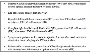
The indications for cardiac resynchronization therapy are clearly supported in the various international heart failure guidelines, with a good degree of evidence 1. Table 1.
Of the previously given indications, the least frequent is resynchronization therapy in patients who develop heart failure due to right ventricular electrical stimulation. However, in some cases, CRT is the only therapeutic alternative which may be offered to improve the ejection fraction and symptoms.
We present the case of a 62-year-old patient with an indication for a dual chamber pacemaker due to complete AV block, who, after implantation of the device, developed heart failure requiring a change of device to a cardiac resynchronizer. This change was necessary since pacemaker induced cardiomyopathy was thought to be the sole cause of heart failure in this patient, after ruling out other more common causes like ischemic heart disease and myocarditis.
Clinical case
The patient is a 62-year-old female with a history of systemic lupus erythematosus, rheumatoid arthritis, and a high-grade atrial ventricular block with dual chamber DDD pacemaker implantation. The studies performed prior to device implantation were as follows: normal echocardiogram, normal cardiac magnetic resonance and heart catheterization without significant coronary artery lesions. She had a 40-day history of decreased functional class, dyspnea with medium to low exertion and lower limb edema. On physical exam she had a blood pressure of 140/70 mm Hg, heart rate of 80 beats per minute and respiratory rate of 18 breaths per minute. Grade II jugular engorgement was seen at 45 degrees, she had a regular heart rhythm with no murmurs, and scant basal respiratory rales. Hepatojugular reflux was present without ascites or hepatomegaly, along with grade II lower limb edema. An electrocardiogram was ordered which showed a pacemaker rhythm (Figure 1), BNP was 4,410 pg/mL and a transthoracic echocardiogram evidenced diminished global systolic function (LVEF:35%) without altered segmental contractility, and left ventricular concentric hypertrophy, greater in the apex. With these findings, she was hospitalized with a diagnostic impression of heart failure with a reduced ejection fraction, and treatment was begun with neurohumoral block and a diuretic.

Figure 1 Electrocardiogram. Pacemaker rhythm. Left bundle branch block morphology. Diffuse repolarization disorder.
Due to her history, she was seen by rheumatology, ruling out lupus activity. A cardiac magnetic resonance (Figure 2) did not show any abnormal enhancements or scarred areas or infiltration suggesting myocarditis or ischemia; there was a decreased ejection fraction of 32% and right chamber dilation. These findings ruled out an ischemic, myocarditis or rheumatic disease etiology, and it was considered to be pacemaker induced cardiomyopathy. Optimal medical treatment for heart failure was continued and the electrical stimulation device was changed from a conventional pacemaker to a cardiac resynchronizer. The patient's symptoms improved, her heart failure was compensated and a subsequent follow up echocardiogram showed a recovered ejection fraction of 50%.
Discussion
Patients with pacemaker stimulated right ventricles may develop various complications, including pacemaker syndrome and pacemaker induced cardiomyopathy. Pacemaker syndrome is an entity which presents in 20% of patients and is an atrioventricular dissociation in single chamber devices programmed in VVI mode, which resolves with implantation of a dual chamber pacemaker with DDD stimulation 3.
Pacemaker induced cardiomyopathy is another common entity which presents with a variable prevalence throughout the use of the device; approximately 19.5% of patients have pacemaker induced cardiomyopathy at three years, and it results from biventricular dyssynchronization due to a high right ventricular stimulation load which leads to a deteriorated left ventricular ejection fraction below 50%, decreasing more than 10% from the patient's baseline 4.
Our patient's LVEF decreased 28%, going from 60-32%, with signs and symptoms of heart failure 12 months after device implantation, which coincides with other studies where an association has been found between pacemaker implantation and the development of cardiomyopathy, even within the first six months of use.
Right ventricular stimulation load is the most important risk factor for ventricular dyssynchrony. The incidence of death and hospitalizations due to heart failure has been reported to reach up to 30% in patients with more than 40% right ventricular stimulation. Our patient initially required only 2% ventricular stimulation; however, she subsequently increased to 98%, increasing the risk of pacemaker induced cardiomyopathy.
The development of heart failure in these patients is not only due to the electromechanical dyssynchronization; rather, this condition is the initial mechanism to which are added oxygen supply-demand imbalance in the myocardium, impaired contractility due to altered filling pressures, cardiac remodeling and the activation of various systems such as the sympathetic nervous system or the renin-angiotensin-aldosterone system which accentuate the signs and symptoms of congestion and/or hypoperfusion even more 4. Ventricular wall hypertrophy (remodeling) was documented in our patient on the echocardiogram and magnetic resonance, which was not seen in the studies prior to device implantation, corresponding with what was previously described. Likewise, medical treatment included beta blockers and angiotensin converter enzyme inhibitors, blocking the various misnamed "compensating" systems.
Among the risk factors for developing pacemaker induced cardiomyopathy, Khurshid et al. found a direct relationship with the male sex (HR 2.15; 95% CI: 1.7-3.94, P < .01) and QRS duration (HR 1.03 for each millisecond increase; 95% CI: 1.01-1.05, P<.001) 5. Lee et al. found a direct relationship to age (HR 1.62; 95% CI: 1.22-2.16, P < .001) 6, and Kiehl et al. to a >20% right ventricular stimulus (HR: 6.76; 95% CI: 2.08-22.0, P < .002) 7. Other risk factors documented in the literature include prior atrial fibrillation and reduced global longitudinal strain following device implantation. In our patient, the main documented risk factor was right heart stimulation, which reached 98%.
Various mechanisms have been used to try to prevent pacemaker induced cardiomyopathy: using a high septal stimulation site instead of apical stimulation has not shown any difference in hard outcomes such as mortality and/or hospitalizations. Perhaps the best strategies which may favor the prevention of cardiomyopathy are choosing an appropriate device, avoiding ventricular overstimulation as much as possible, and immediate implantation of cardiac resynchronization devices in patients with already established heart failure. Curtis et al. showed that dual chamber stimulation in patients with an ejection fraction below 50% decreased mortality and hospitalizations due to heart failure when compared with patients with only right ventricular stimulation (HR 0.74 95% CI 0.60-0.90) 8. Our patient had no prior deterioration of the ejection fraction, and therefore dual chamber stimulation was not initially considered, but the development of heart failure made a change of cardiac stimulation device necessary.
Gage et al. showed how a change from a single chamber pacemaker to dual chamber stimulation (resynchronization therapy) is related to an improved ejection fraction (8.3 ± 9 vs. 5.8 ± 9, P =0.005) and a lower risk of death or hospitalizations due to heart failure (HR 0.67 95% CI 0.51-0.89, P =0.005) 9.
Studies in patients with a preserved ejection fraction have also been conducted. Man Yu et al. reported that at 12 months follow up, there was far less deterioration in the ejection fraction of patients with biventricular stimulation than those with right ventricular stimulation (54.8% vs. 62.2, P<0.001) 10. Studies with a longer follow up period have also been carried out, showing a smaller reduction in ejection fraction and end systolic volume after two years of biventricular stimulation 11. However, we must take into account that the costs of resynchronization therapy are much higher, which would not be cost effective in our population, and the procedural complications are also greater compared to conventional pacemaker implantation 12. These are perhaps the reasons why a resynchronizer was not initially implanted in our patient.
In this patient, a three-month waiting period to determine the therapeutic response to medications was not observed since the cause of heart failure was clearly established. Thus, pacemaker induced cardiomyopathy could be one of the conditions in which the patient could benefit from CRT without completing three months of optimal medical management 13.
After the device change, the patient showed a significant clinical improvement, which had not been achieved even with the optimal tolerated medical treatment. To date, she continues to be free of cardiovascular symptoms, has no clinical signs of congestion or low cardiac output, and has not had any readmissions for heart failure.
Conclusion
Complications associated with electrical stimulation devices should be considered as one of the common causes of heart failure. The prompt identification of these complications and replacement with biventricular stimulation devices improve the hemodynamic parameters such as ejection fraction, and decrease the symptoms, number of hospitalizations and risk of death in these patients.


 text in


