Introduction
Coronary artery aneurysms are a rare disorder with a variable reported frequency ranging from 0.9 to 4.9% 1. They are defined as coronary dilations which are more than 1.5 times the diameter of adjacent segments 2, and they are classified according to their diameter: if the transverse diameter is greater than the longitudinal diameter they are known as "saccular", and if the longitudinal diameter is greater than the transverse, they are known as "fusiform" 3. The most common location is in the right coronary, and the least frequent is in the left common trunk 4,5.
The average age at which this pathology is identified is 69 years, and it is more frequent in people over the age of 70, with a higher prevalence in males. The main associated risk factors have been diabetes mellitus, current smoking, and hyperlipidemia, with the latter and male sex having the highest statistical significance 5,6. Its etiology correlates with the age at presentation: in young patients, the most common etiologies are Kawasaki disease, hyperhomocysteinemia, connective tissue disorders and congenital disorders; in patients over the age of 40, the main cause is atherosclerosis 7. Other reported causes include mycoses and the use of alkaloids such as cocaine 2,7.
The pathophysiology of coronary aneurysms is not well known; it is thought that an increased inflammatory response in the vascular wall and the activation of metalloproteinases may be contributing factors 2. Significant coronary lesions (greater than 70% occlusions) associated with this pathology are common (75.6%), according to the findings of a study of 978 diagnosed cases of coronary aneurysms 1. Another, smaller, study reported that multivessel disease may even be present 5. Likewise, Baman et al. reported the presence of coronary aneurysms as an independent adverse effect for long-term mortality 6.
Case description
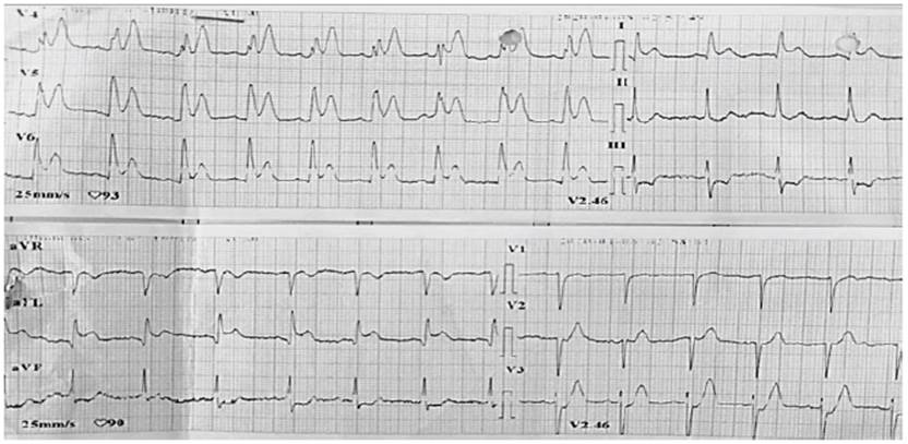
This was a 25-year-old male patient with no reported past medical, toxicological or surgical history, with class II obesity (body mass index 37 kg/m2), who presented to the emergency room due to 20 minutes of oppressive chest pain radiating to the left arm, neck and jaw, accompanied by diaphoresis and a feeling of dyspnea. On the admission physical exam, he was found to have arterial hypertension (140/90 mmHg), with no pathological findings on cardiopulmonary auscultation. A twelve-lead electrocardiogram showed sinus rhythm, a heart rate of 90 beats per minute, and ST elevation in leads DI, AVL, V4 and V5 (Figure 1).
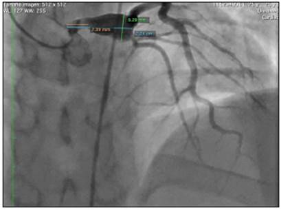
An emergency coronary angiography was performed, revealing an aneurysm in the main trunk of the left coronary artery measuring 9.29 mm at its widest point, with the normal portion of the vessel measuring 3.76 mm. There was also a thrombus at this point occluding the anterior descending artery, no significant angiographic lesions in any of the other coronary arteries, and TIMI 3 distal flow (Figure 2). Coronary angioplasty was attempted, but the anterior descending occlusion could not be passed; therefore, tirofiban was begun for 24 hours. A transthoracic echocardiogram documented the existence of extensive anteroseptal apical akinesia, and moderate global left ventricular dysfunction with a 32% ejection fraction.

Figure 2 Coronary angiography image registering coronary trunk measurements of ap proximately 22 mm, proximal portion of the vessel 7.39 mm wide and aneurysmal segment approximately 9.29 mm wide.
He was transferred to the intensive care unit, and during his hospital stay he developed acute decompensation with cardiogenic shock which required vasopressor support with noradrenaline and inotropic support with levosimendan. After hemodynamic compensation, follow up coronary angiography was performed which showed that the thrombus had disappeared, and the same coronary anatomy characteristics persisted.
Due to the anatomical location of the aneurysm, the option of surgical revascularization was considered. However, the dobutamine stress echocardiography stratification showed a 32% ejection fraction, with infarction and a non-viable anterior wall, and therefore continued medical management was ordered.
He was discharged on double antiaggregation therapy with acetylsalicylic acid 100 mg/day and clopidogrel 75 mg/day, which was discontinued at a follow up visit after the acute event, at which time antithrombotic treatment with rivaroxaban 2.5 mg q 12 hours was added. After six months of follow up he has had no further clinical outcomes.
Discussion
Coronary artery aneurysms are a rare disorder which present predominantly in older adults, making our case more interesting, as it occurred in a young adult. Furthermore, according to the literature review performed, these aneurysms seem to form preferentially in the right coronary; in two of the studies with the greatest number of cases, the right coronary was the number one reported location (proximal and middle segment), followed by the anterior descending and circumflex 1,4 and, finally, the main trunk of the left coronary, with a frequency as low as 0.1%, as reported by Topaz et al. in a review of 22,000 angiographies 8, which gives a special connotation to our case. Other studies also report the right coronary in first place, followed by the circumflex and, finally, the anterior descending 9.
With regard to coronary aneurysm treatment, surgical revascularization procedures have been reported in some cases with grafts to the coronaries 9. Authors such as Kawsara et al. propose a management algorithm (Figure 3), but there is no consensus due to the incomplete understanding of its pathophysiology and lack of sufficient studies with this type of population to validate its use. Therefore, management should be focused on each patient, individually, depending on how the disease manifests, and the location and morphology of the aneurysm 10.

Figure 3 Suggested algorithm for management of coronary artery aneurysms. Modified from Kawsara et al.
The presented case represents a therapeutic challenge, considering that the patient debuted with a thrombus in the aneurysmal dilation of the coronary trunk, leading to an ST-elevation acute myocardial infarction with no residual viability of the anterior wall, thus ruling out surgical treatment as a therapeutic option. In the end, secondary prevention with antiplatelet treatment and the subsequent addition of antithrombotics was ordered, despite the limited evidence in this population.
Conclusion
Coronary aneurysms are a rare entity, with little scientific evidence, and thus there is no consensus on their management. Large studies are needed to determine the use of anticoagulant or antiplatelet therapy. For now, it is important for each case to be evaluated individually, considering location, anatomical morphology, clinical presentation and myocardial involvement, among other things.


 text in


