SciELO - Scientific Electronic Library Online

 
vol.46 issue120New records, validated name, and redefinition in Azolla (Salviniaceae, Pteridophyta) from Costa RicaBioreduction in vitro of hexavalent chromium using a microbial consortium author indexsubject indexarticles search
Home Pagealphabetic serial listing  

Services on Demand

Journal

Article

Indicators

Related links

  • On index processCited by Google
  • Have no similar articlesSimilars in SciELO
  • On index processSimilars in Google

Share


Actualidades Biológicas

Print version ISSN 0304-3584

Actu Biol vol.46 no.120 Medellín Jan./June 2024  Epub Aug 22, 2024

https://doi.org/10.17533/udea.acbi/v46n120a03 

Short communication

First record of an ocular anomaly in Carollia perspicillata (Chiroptera: Phyllostomidae) in the Republic of Panama

Primer registro de anomalía ocular en Carollia perspicillata (Chiroptera: Phyllostomidae) en la República de Panamá

Nelson Guevara-Alvarado1  2  * 
http://orcid.org/0000-0002-3200-6648

Yulissa Díaz-Hernández1 
http://orcid.org/0009-0009-0659-3250

¹ Biomundi Foundation, Panama City, Panama.

² College of Biologists of Panama, COBIOPA, Panama City, Panama.


ABSTRACT

Ocular anomalies caused by injury, disease or genetic mutations result in total or partial loss of vision, which affects the normal development and behavior of affected individuals in their natural environment. In bats, these cases are rarely reported. In addition, it is possible that researchers occasionally encounter these anomalies, but they are not reported due to various factors. This paper documents, for the first time, an individual of Carollia perspicillata with an ocular anomaly (ocular inflammation) in the Republic of Panama, recorded in the Bunorgandi Private Nature Reserve in the Province of Panama.

Keywords: bats; Carolliinae; ocular inflammation; ocular lesions

RESUMEN

Las anomalías oculares causadas debido a lesiones, enfermedades o mutaciones genéticas generan la pérdida total o parcial de la visión, lo cual afecta el desarrollo y comportamiento normal de los individuos afectados en su entorno natural. En murciélagos, estos casos raramente son reportados. Además, es posible que en ocasiones los investigadores encuentren estas anomalías, pero no son reportadas debido a diversos factores. En este trabajo se documenta por primera vez a un individuo de Carollia perspicillata con anomalía ocular (inflamación ocular) en la República de Panamá, registrado en la Reserva Natural Privada Bunorgandi de la Provincia de Panamá.

Palabras clave: Carolliinae; inflamación ocular; lesión ocular; murciélagos

Although bats mainly use echolocation to move in their environment, they can use signals for orientation and movement (Suthers and Wallis, 1970), so visual organs are highly functional, even if they are small and with limited vision (Müller et al., 2009). It has been reported that, during the search for food and new refuges, vision aids in predator avoidance and, in some species, in the capture of prey (Altringham and Fenton, 2003; Eklöf and Jones, 2003), including the detection of flowers that reflect U.V. rays for those that feed on nectar (Winter et al., 2003).

Ocular abnormalities in bats have been scarcely reported (Sánchez-Hernández et al., 2019). Some of the ocular lesions observed include corneal opacity, anophthalmia, microphthalmia, and bacterial infections (Sánchez-Hernández et al., 2019). Recently, Turner et al. (2021) listed a series of ocular diseases in species of the family Pteropodidae, where corneal opacity leads, followed by cataracts, anterior uveitis, adnexal diseases and glaucoma. The most common causes of these diseases are trauma from interindividual and intraindividual aggression and falls or shocks during flight, which may be linked to underlying diseases that cause weakness (Turner et al., 2021). Other causes of these diseases are genetic predisposition, degenerations and environmental factors (Davidson and Nelms, 2013). Even in a study of ocular lesions in mammals, it is evident that the rabies virus extends to the eye, possibly through the ocular nerve (Dalton et al., 2020). Therefore, this would be another cause of ocular abnormalities in bats, as they may be reservoirs of the rabies virus (Scheffer et al., 2014).

The infrequency and scarcity of reports on the subject may indicate that it is not given the required importance or that cases go unnoticed for various reasons such as the small size of the eyes of some species (Sánchez-Hernández et al., 2019). The importance of reporting these anomalies in bats, whether in their eyes or another part of the body, marks a milestone in terms of studies on mortality due to diseases and their possible causes, and in turn is of utmost relevance for the study of ecosystem health (Bayon et al., 1999; Turner et al., 2021).

The objective of this report is to present the first case of ocular anomaly in Carollia perspicillata (Linnaeus, 1758) in the Republic of Panama. Since it is a product of an unexpected observation during the development of the main study and we do not have the necessary materials for an adequate sample collection for any veterinary analysis, we cannot hypothesize about possible pathological or genetic causes of the anomaly.

On February 9, 2023 in the Bunorgandi Private Nature Reserve (Figure 1) located in the Corregimiento de Cañitas, Chepo District, Province of Panama, Republic of Panama (9˚14̍ 53̎ N 78˚45̍ 09̎ W) of 53 individuals and 18 species, an adult male individual of C. perspicillata that presented an ocular anomaly was captured (Figure 2). The individual was captured at 19:55 hours using mist nets and identified using the guidelines of Reid (2009) and York et al. (2019). The individual presented swelling of the right eye globule and a small yellow coloration within the eye globule, which we assumed to be sightless as it did not respond to visual stimuli such as white and yellow light from flashlights, and proximity of objects. However, the left eyeball presented apparent normality, functional vision and responded to visual stimuli. Once the individual's data was recorded, it was released near the capture zone.
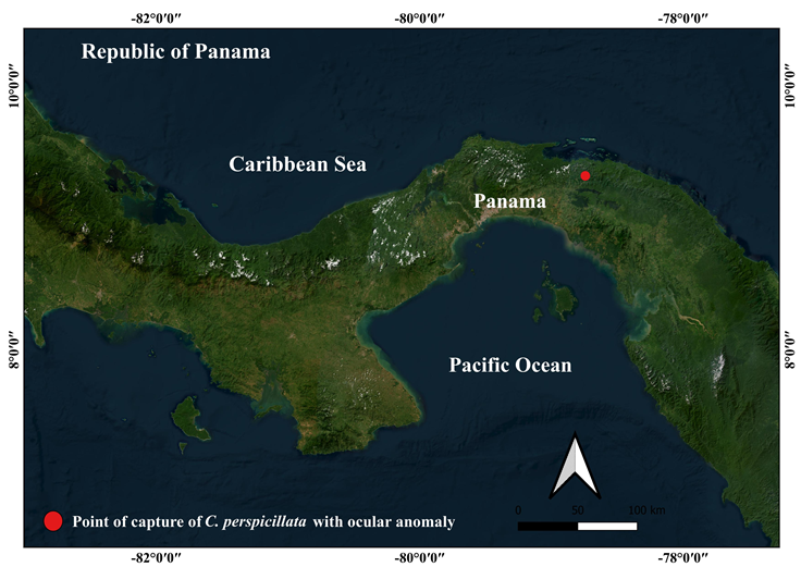
Figure 1 Capture area of C. perspicillata with ocular anomaly in the Bunorgandi Private Nature Reserve, Republic of Panama. 
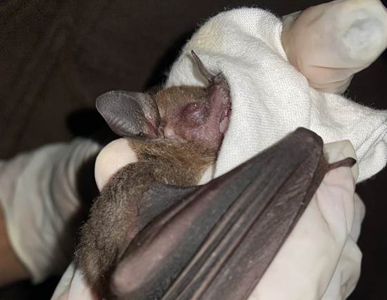
Figure 2 Individual of C. perspicillata captured in the Bunorgandi Private Nature Reserve with ocular anomaly in the right eyeball. 

There are few records of ocular lesions, anomalies or diseases in bats, currently restricted to a few species in only five reports (Table 1).

Table 1 List of ocular lesions, anomalies or diseases recorded in bats. 

Species Eye injuries or diseases Country Reference
Yangochiroptera
Eptesicus fuscus Closed eyelids, unpigmented choroidal layer, undifferentiated retina and underdeveloped crystalline lens. United States Kunz and Chase (1983)
Carollia perspicillata Blindness. Brazil Feijó et al. (2018).
Artibeus planirostris Corneal opacity.
Artibeus lituratus
Artibeus jamaicensis Corneal opacity, ocular injury. Mexico Sánchez-Hernández et al. (2019).
A. lituratus Corneal opacity, Active infection with subconjunctival hemorrhage, Anophthalmia.
Desmodus rotundus Ocular infection.
Glossophaga soricina Anophthalmia.
Sturnira parvidens Ocular injuries with corneal perforation in the eye.
Tadarida brasiliensis Microphthalmia or nanophthalmia.
T. brasiliensis Ocular injury (non-specific). United States Dalton et al. (2020)
Myotis lucifugus United States
Yinpterochiroptera
Pteropus vampyrus Lens dislocation, cataracts, glaucoma, anterior uveitis, corneal disease, adnexal diseases (conjunctivitis, blepharitis, meibomianitis and partial tear of the lateral rectus muscle) and orbital diseases (retro bulbar abscesses, orbital fractures and acute traumatic proptosis. United States Turner et al. (2021)
P. pumilus
P. hypomelanus
P. giganteus
P. rodricensis
Cynopterus brachyotis
Rousettus aegyptiacus

The little information available on eye injuries and diseases in bats generates several questions about how the lack of vision, which is complementary to echolocation signals (Feijó et al., 2018), affects the species in the wild to fulfill their functions of orientation, feeding and movement at short and long distances (LaVal and Rodríguez, 2002). Therefore, promoting reports of this type helps to gather information that can allow generating a basis for future studies more focused on analyzing the causes and consequences of ocular anomalies, injuries and diseases.

Acknowledgments

We thank the Biomundi Foundation, the Bunorgandi Private Nature Reserve and Renata Sponer for the organization and logistical support. To the Biology students of the University of Panama Deyvis Castillo, Cristian Guzmán, Luis Jiménez and Esthefany Izasa for their support in the collection and field data collection. The English version was translated by Actualidades Biólogicas Journal.

References

Altringham, J. D., & Fenton, M. B. (2003). Sensory Ecology and Communication in the Chiroptera. Bat Ecology (pp. 90-127). Chicago, Estados Unidos: University of Chicago Press. [ Links ]

Bayon, A., Brotons, N. J., Albert, A., & Talavera, J. (1999). Patología ocular en reptiles. Clínica Veterinaria de pequeños Animales (AVEPA), 19(4), 227-242. https://ddd.uab.cat/record/68645Links ]

Dalton, M. F., Siepker, C. L., Maboni, G., Sanchez, S., & Rissi, D. R. (2020). Ocular and lacrimal gland lesions in naturally occurring rabies of domestic and wild mammals. Veterinary Pathology, 57(3), 409-417. https://doi.org/10.1177/0300985820911458 [ Links ]

Davidson, M. G., & Nelms, S. R. (2013). Diseases of the lens and cataract formation. Veterinary Ophthalmology (pp. 1199-1233). New Jersey, Estados Unidos: Wiley-Blackwell. [ Links ]

Eklöf, J., & Jones, G. (2003). Use of vision in prey detection by brown long-eared bats, Plecotus auritus. Animal Behaviour, 66(5), 949-953. https://doi.org/10.1006/anbe.2003.2272 [ Links ]

Feijó, A., Hannah, N., Emmanuel, V. M., & da Rocha, P. A. (2018) Blindness in echolocating bats. Mammalia, 83(3), 272-275. https://doi.org/10.1515/mammalia-2018-0034 [ Links ]

Kunz, T. H., & Chase, J. (1983). Osteological and ocular anomalies in juvenil big brown bats (Eptesicus fuscus). Canadian Journal of Zoology, 61, 365-369. https://doi.org/10.1139/z83-048 [ Links ]

LaVal, R., & Bernal, R. (2002). Murciélagos de Costa Rica Bats. Costa Rica: Editorial INBio. [ Links ]

Müller, B., Glösmann, M., Peichl, L., Knop, G. C., Hagemann, C., & Ammermüller, J. (2009). Bat Eyes Have Ultraviolet-Sensitive Cone Photoreceptors. PLoS ONE, 4(7), e6390. https://doi.org/10.1371/journal.pone.0006390 [ Links ]

Sánchez-Hernández, C., Silvia S., Guerrero, S., Romero-Almaraz, M., Sil-Berra, L., & Schnell, G. D. (2019). Ocular Lesions and Diseases in Bats from Jalisco and Oaxaca, Mexico. Acta Chiropterologica, 20(2), 485-492. https://doi.org/10.3161/15081109ACC2018.20.2.018 [ Links ]

Scheffer, C. K., Iamamoto, K., Asano, M. K., Mori, E., Garcia, E. A. I., Achkar, S. M. (2014). Murciélagos hematófagos como reservorios de la rabia. Revista Peruana de Medicina Experimental y Salud Publica, 31(2), 302-9. http://www.scielo.org.pe/scielo.php?script=sci_arttext&pid=S1726-46342014000200018Links ]

Suthers, R. A., & Wallis, N. E. (1970). Optics of the eyes of echolocating bats. Vision Research, 10(11), 1165-1173. https://doi.org/10.1016/0042-6989(70)90034-9 [ Links ]

Turner, R. C., Alexander, A. B., Wellehan, J. F. X., Heard, D., Abbott, J. R., Crevasse, S. E., & Plummer, C. E. (2021). Retrospective analysis of ocular disease in a population of captive pteropodid bats, 2003-2020. Veterinary Ophthalmology, 24(3), 240-251. https://doi.org/10.1111/vop.12879 [ Links ]

Reid, F. A. (2009), A field guide of the mammals of Central American & southeast Mexico. New York, Estados Unidos: University of Chicago Press. [ Links ]

York, H. A., Rodríguez-Herrera, B., LaVal, R., & Timm, R. (2019). Field keys to the bats of Costa Rica and Nicaragua. Journal of Mammalogy, 100, 1726-1749. https://doi.org/10.1093/jmammal/gyz150 [ Links ]

Winter, Y., López, J., & Von Helversen, O. (2003). Ultraviolet vision in a bat. Nature, 425, 612-614. https://doi.org/10.1038/nature01971 [ Links ]

Author Contributions Nelson Guevara-Alvarado participated in the data collection, search for information, preparation of the map, writing and correction of the manuscript. Yulissa Díaz-Hernández, participated in the data collection, search for information, preparation of the introduction, Table 1, writing and proofreading of the manuscript.

Received: February 28, 2023; Accepted: October 12, 2023

*Corresponding author: nelson2295@hotmail.com

Conflict of Interest

There are no conflicts of interest.

Creative Commons License This is an open-access article distributed under the terms of the Creative Commons Attribution License