Services on Demand
Journal
Article
Indicators
-
 Cited by SciELO
 -
 Access statistics
Related links
-
 Cited by Google
 -
 Similars in
SciELO
 -
 Similars in Google
Share
Caldasia
Print version ISSN 0366-5232On-line version ISSN 2357-3759
Caldasia vol.29 no.1 Bogotá Jan./June 2007
EPIDERMAL CHARACTERS OF BACCHARIS(ASTERACEAE) SPECIES USED IN TRADITIONAL MEDICINE
Caracteres epidérmicos de las especies del género Baccharis (Asteraceae) usadas en la medicina popular
SUSANA E. FREIRE
ESTRELLA URTUBEY
DANIEL A. GIULIANO
División Plantas Vasculares, Museo de La Plata, Paseo del Bosque, 1900 La Plata, Argentina. freire@museo.fcnym.unlp.edu.ar
ABSTRACT
A morphological study of 38 species of Baccharis used in traditional medicine was carried out to provide some epidermal characters that will contribute to the knowledge of the genus. The present study revealed: 1) seven different types of trichomes: conical, aseptate flagellate, filiform flagellate, 1-armed, 2-4-armed, bulbiferous flagellate, and glandular biseriate; 2) that 28 of the total of 38 species have trichomes in tufts; 3) six different types of stomata: anomocytic, anisocytic, cyclocytic, actinocytic, tetracytic, and staurocytic; 4) that some trichome types, such as 2-4-armed (B. dracunculifolia) and aseptate flagellate branched (B. trinervis), show a high diagnostic value; 5) that the stomata types can be used to differentiate species with similar trichomes type (e.g. B. trimera and B. articulata). Illustrations of the studied characters are provided.
Key words. Baccharis, Compositae, medicinal species, stomata, trichomes.
RESUMEN
Se realizó un estudio de caracteres epidérmicos de las hojas de las 38 especies de Baccharis usadas en la medicina popular. El análisis de los caracteres revela: 1) siete tipos de tricomas: cónicos, flageliforme aseptados, filiformes flagelados, 1-armados, 2-4-armados, bulbíferos flagelados y glandulares biseriados; 2) que 28 especies del total de 38 especies medicinales estudiadas presentan tricomas en “nidos pilosos”; 3) que seis tipos de estomas están presentes: anomocíticos, anisocíticos, ciclocíticos, actinocíticos, tetracíticos y estaurocíticos; 4) que algunos tricomas muestran un alto valor diagnóstico, por ejemplo, los tricomas 2-4-armados son exclusivos de B. dracunculifolia y los flagelados-aseptados ramificados están presentes sólo en B. trinervis; 5) que los tipos de estomas permiten la diferenciación de especies con igual tipo de tricoma, (por ejemplo B. trimera de B. articulata). Se incluyen ilustraciones de los caracteres diagnósticos.
Palabras clave. Baccharis, Compositae, especies medicinales, aparatos entomáticos, tricomas.
INTRODUCCIÓN
Baccharis L. is one of the largest genera of the tribe Astereae (Nesom, 1994). It comprises c. 400 American species of shrubs or subshrubs, occasionally small trees and herbs, nearly all dioecious.
According to Zin (1922), Amat (1983), Zardini (1984), Girault (1987), Boldt (1989), Correa & Bernal (1990), Iharlegui & Hurrell (1992), Soria (1993), Heinrich (1996), Rojas et al. (1999), Pérez-García et al. (2001), Erazo et al. (2002), Baggio et al. (2003), Vidari et al. (2003), Souza (2004), and a recent webpage (Harding-Barlow), 38 medicinal species of Baccharis with folk medicinal use (or mentioned at least as medicinal herbs) are recognized (Table 1); many of these species can be distinguished by their leaf or winged stem morphology (Appendix 1). In 22 of these species biological activity was tested (Gutkind et al. 1981, Vidari et al. 2003, Verdi et al. 2005). Baccharis megapotamica and B. pedunculata are also included in this study since they can be considered promisory medicinal species judging from their tested biological activity (Verdi et al. 2005).
Appendix 1. Baccharis species used in traditional medicine.
Heering (1899), Volkens (1890), Quentin (1911), Ariza Espinar (1973), Barroso (1976), Pertusi (1987), Hellwig (1990, 1992), Müller (2006) and, are among authors who have contributed most to the solving taxonomic problems through the analysis of micromorphology of leaf surface in Baccharis.
Epidermal traits, i.e. epidermal cells, stomata, and hairs, have proven to be an important tool in taxa delimitation in many plant families (e.g. Lackey 1978, Metcalfe & Chalk 1950-1979, Sinclair & Sharma 1971, Uphof et al. 1962), and also in distinguishing medicinal species since drugs of pharmaceutical use are made up of dried and bruised parts in which the different macroscopical characteristics of the species are not generally distinguishable (e.g. Amat, 1988; Rapisarda et al., 1997). Within this context, this study pretend to contribute to the knowledge of the medicinal species of the genus Baccharis.
MATERIAL AND METHODS
The study was performed using dried leaves or winged stems (if only bract-like leaves are present) taken from herbarium specimens (Appendix 2). Accepted names, synonyms, vernacular names, distribution, medicinal uses, and biological activity for each studied species are given in Table 1.
Appendix 2. Representative specimens examined of Baccharis (one specimen per species is cited).
The epidermal microcharacters were studied in samples cleared using the technique of Dizeo de Strittmatter (1973), and stained ussing safranin in 80% ethanol. Measurements of stomata (length) were taken using a Nikon light microscope equipped with an ocular micrometer. The average size of stomata was determined based on measurements performed on 15-20 replicates per sample.
Descriptive terminology for the trichomes basically follows Ramayya (1962). Whenever possible, additional synonyms of trichome terminology were added (Ariza Espinar 1973, Metcalfe & Chalk 1989, Müller 2006). The classification of anticlinal epidermal cell wall patterns follows Stace (1965). Stomata types were classified according to Stace (1965), Van Cotthem (1970), Metcalfe & Chalk (1979). The nomenclature of the included species follows Matuda (1957), Cuatrecasas (1967), Ariza Espinar (1973), Barroso (1976), Giuliano (2000) and Oliveira et al. (2006). Drawings were made by the authors using a microscope Leitz SM Lux with camera lucida.
RESULTS AND DISCUSSION
Epidermal characters (Table 2)
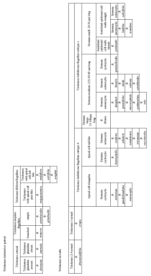
Leaf pubescence. All studied species of Baccharis are pubescent (only a few, e.g. B. dracunculifolia and B. trinervis are subglabrous at maturity). Two major groups of indumentum can be distinguished within the medicinal species, one consisting of isolated trichomes and the other with trichomes in tufts (= “nidos pilosos”, Ariza Espinar, 1973; = “Haarnester”, Hellwig 1992).
Tabla 2. Epidermal characters.
Seven different types of trichomes were found:
(1) Conical trichomes: uniseriate, 5-6-celled with body gradually narrowed to a sharp point. Cells longer distally, often nodulose at the joints. Lateral and cross walls slightly thickened. This trichome type is present in B. anomala and B. decussata. (Fig. 1 A). Similiar trichomes were illustrated by Müller (2006) for B. decussata subsp. jelskii (sub “pedestal hair”) and by Ariza Espinar (1973) for the non-medicinal B. pulchella.
Figura 1. Trichomes.- A-E, Isolated trichomes. A: conical, B. anomala, B: aseptate flagellate branched, B. trinervis; C and D, Filiform flagellate. C: B. multiflora; D: B. megapotamica; E: aseptate flagellate simple, B. artemisioides; F-L, Tufted trichomes (“pilose nest”) of eglandular trichomes and biseriate glandular trichomes. F: 1-armed trichome, B. crispa; G: 2-4-armed trichomes, B. dracunculifolia; H: bulbiferous flagellate, subtype a, B. boliviensis; I and J, Bulbiferous flagellate, subtype b. I: B. notosergila; J: B. teindalensis; K and L, Bulbiferous flagellate, subtype c. K: B. pedunculata; L: B. tola.
(2) Aseptate flagellate trichomes (or whip trichomes, Metcalfe & Chalk 1989): uniseriate, 2-3-celled with body differentiated in stalk and a long, whip-like terminal cell. Stalk 1-2-celled, cells usually isodiametrical or broader than long.
(2) (a) Simple: terminal cell very long, flagellate, tubular, as wide as the cells of the stalk. This trichome type is present in B. artemisioides and B. grisebachii (Fig. 1 E), which have discolorous leaves with white-lanate abaxial surface. Trichomes of B. artemisioides were previously analyzed and illustrated by Ariza Espinar (1973) and Pertusi (1987). The aseptate flagellate trichomes of B. grisebachii were illustrated by Hellwig (1992) and by Müller (2006), sub “flagellate filiform” and “filiform hair” respectively.
(2) (b) Branched: three to five whip trichomes (with stalk 1-4-celled) appearing stellately branched from the only stalk cell. This trichomes type is present isolated only in B. trinervis (Fig. 1 B).
(3) Filiform flagellate trichomes: uniseriate, many-celled, body filiform to cilindrical and slightly tapering above. Cells usually broader than long, isodiametrical, or slightly longer than broad, with lateral walls convex. Terminal cell relatively short, tail-like and pointed (B. multiflora, B. serrifolia and B. wrightii, in the latter the trichomes are only found on the stem, Fig. 1 C) or pear-like and rounded at the apex (B. megapotamica, Fig. 1 D); those with tail-like apical cell can be straight or incurved. Dense contents have been seen in the terminal cell of these trichomes, which probably have secretory function.
(4) 1-armed trichomes: uniseriate, 3-4-celled, with body differentiated into stalk and head. Stalk 2-3-celled, cells usually broader than long, lateral walls straight. Head 1-celled (occasionally branched at the base), with thick lateral and cross walls and pointed or rounded at the apex. This trichome type is present forming tufts or pilose nests only in B. crispa (Fig. 1 F). It was previously analyzed and illustrated by Ariza Espinar (1973) for B. crispa. Hellwig (1992) found trichomes 1-armed in a non-medicinal species, B. rhomboidalis and also in B. macraei and B. pilcensis (Hellwig, 1990).
(5) 2-4-armed trichomes: uniseriate, 4-5-celled, with body differentiated into stalk and head. Stalk 3-4-celled, cells broader than long with lateral walls convex. Head 1-celled, cell 2, 3 or 4-branched (falls/collapsing at maturity). This trichome type is present forming tufts or pilose nests (associated or not to glandular trichomes) only in B. dracunculifolia. (Fig. 1 G). Müller (2006) described and illustrated these trichomes of Baccharis dracunculifolia as “uniseriate hairs with branched terminal cells”. Hellwig (1992) found 4-armed trichomes in a non-medicinal species B. erioclada.
(6) Bulbiferous flagellate trichomes: uniseriate, 5-6-celled, usually forming tufts or pilose nests with glandular trichomes. According to the number of subapical cells and the lenght of the apical cell, three subtypes can be distinguished:
Subtype a: body differentiated into stalk and head. Stalk 2-3-celled, cells slightly longer than broad or isodiametrical, terminal cells of the stalk 2, swollen, sphaerical or oblong-ovoid in shape. Head 1-celled, long, flagellate. This trichome type is present only in B. boliviensis. (Fig. 1 H). Similar trichomes are also present in the unrelated non-medicinal B. pingraea (Hellwig, 1992).
Subtype b: body with cells usually enlarging above, resulting cuneate in shape. Terminal cell short with dense contents, broadly triangular (broadly triangular in B. notosergila or narrowly triangular in B. articulata, B. gaudichaudiana, and B. trimera) or more often tail-like, i.e. sharply delimited from the subapical cell (B. conferta, B. heterophylla, B. pilularis, B. teindalensis, B. tricuneata, B. vaccinioides). (Fig. 1 I, J). Trichomes of B. articulata and B. trimera were previously analyzed and illustrated by Pertusi (1987). Hellwig (1992) found trichomes with tail-like apical cell in the unrelated non-medicinal B. paniculata.
Subtype c: this type represents a modification of the subtype a with a differentiated head constituted by a flagellate cell, equal or longer than body, tapering above. This trichome type is present as isolated trichomes in three species, B. anomala, B. coridifolia, and B. trimera, and forms tufts or pilose nests in 16 of the 38 studied species. (Fig. 1 K, L). Bulbiferous trichomes with flagellate apical cell of the medicinal species B. linearis and B. salicifolia were previously illustrated by Ariza Espinar (1973) and Hellwig (1992) respectively. Müller (2006) illustrated for B. coridifolia two types of trichomes, i.e. bulbiferous flagellate subtype b (sub “uniseriate hairs”) and aseptate flagellate (sub “filiform hairs”).
(7) Biseriate glandular trichomes: such trichomes are constituted by 2 rows of cells in the body. Biseriate glandular trichomes are widespread in many taxa studied. They form tufts with non-glandular trichomes; occasionally, tufts are constituted exclusively by two or three glandular trichomes (e.g. B. vaccinioides).
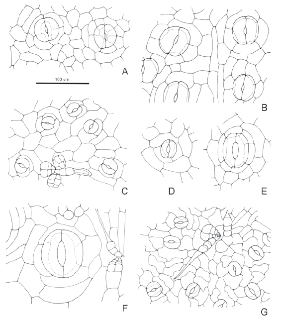
Stomata (Fig. 2): eleven species (B. boliviensis, B. conferta, B. linearis, B. nitida, B. odorata, B. pilularis, B. salicifolia, B. sarothroides, B. teindalensis, B. tola, and B. tricuneata) have actinocytic stomata (Fig. 2 A, B), with five to seven subsidiary cells radially. One species (B. crispa) has anisocytic stomata (Fig. 2 C), with three subsidiary cells, of which one is smaller than the other two. Six species (B. articulata, B. gaudichaudiana, B. genistelloides, B. heterophylla, B. illinita, and B. notosergila) have cyclocytic stomata (Fig. 2 E, F), with four to seven subsidiary cells forming a narrow ring. In three species (B. boliviensis, B. conferta, B. salicifolia) with predominantly actinocytic stomata, a few tetracytic stomata are present (Fig. 2 B), which have four subsidiary cells, two lateral and two polar. Only one of this species (B. conferta) has also staurocytic stomata (Fig. 2 A), with four subsidiary cells with anticlinal walls arranged crosswise to its guard cells. The remaining species have anomocytic stomata (Fig. 2 D, G).
Figura 2. Stomata.- A: actinocytic and staurocytic stomata, B. conferta; B: actinocytic and tetracytic stomata, B. boliviensis; C: anisocytic stomata, B. crispa; D: anomocytic stomata, B. trimera; E: cyclocytic stomata, B. notosergila; F: cyclocytic stomata, B. illinita; G: anomocytic stomata, B. pentlandii.
Cyclocytic stomata in B. articulata, anisocytic stomata in B. trimera, and anomocytic stomata in B. artemisioides were previously reported by Pertusi (1987). Ariza Espinar (1973) analyzed and illustrated actinocytic stomata in B. salicifolia (anomocytic sensu Ariza Espinar), anomocytic stomata in B. coridifolia, and anisocytic stomata in B. crispa (anomocytic sensu Ariza Espinar).
The majority of the species have stomata between 20 to 60 µm long. In only three species the stomata are more than 60 µm long, i.e. Baccharis articulata and B. gaudichaudiana, between 60 to 75 µm long, and B. illinita with stomata between 75 to 105 µm long.
Most of the species analyzed have amphistomatic leaves, only twelve species, i.e. Baccharis anomala, B. decussata, B. dracunculifolia, B. grisebachii, B. heterophylla, B. illinita, B. latifolia, B. megapotamica, B. multiflora, B. pedunculata, B. serrifolia, and B. trinervis.
Anticlinal epidermal cell walls (on abaxial surface): only three species (B. anomala, B. decussata, and B. pentlandii) amongst the medicinal species, have sinuate anticlinal wall (Stace’s types 5-6). The remaining species have straight to slightly undulate anticlinal walls (Stace’s types 1-2).
CONCLUSIONS
Certain anatomical characters allow to the distinction of medicinal species of Baccharis. For example: 1) B. trinervis is recognized from other medicinal species by its branched aseptate flagellate trichomes that are unique within the whole genus; 2) filiform trichomes with pear-like apical cell are exclusive of B. megapotamica; 3) only two species, among medicinal species, B. artemisioides and B. grisebachii, have aseptate flagellate trichomes; 4) B. boliviensis and B. dracunculifolia can be recognized among medicinal species by its bulbiferous trichomes with two subapical cell and 2-4-armed trichomes, respectively; 5) B. illinita is distinguished from other medicinal species by its large stomata of 75-100 µm.
Other characters, such as stomata type and epidermal cell walls, can be used to differentiate species with similar type of trichomes, as a secondary feature. In B. articulata, B. gaudichaudiana, B. notosergila, and B. trimera, the trichomes are bubiferous flagellate with triangular apical cell; however, B. trimera can be distinguished by its anisocytic stomata. In B. heterophylla the trichomes are bulbiferous flagellate with tail-like apical cell, as well as in B. conferta, B. pilularis, B. teindalensis, B. tricuneata, and B. vaccinioides; however, B. heterophylla can be differentiated from them by its cyclocytic stomata. Baccharis pentlandii has bulbiferous flagellate trichomes with long apical cell, which is a common type within medicinal species, but it can be distinguished by its sinuate anticlinal epidermal cell walls on abaxial surface.
ACKNOWLEDGEMENTS
Thanks are given to Jochen Müller and two anonimous reviewers for their valuable comments on the manuscript. Special thanks to the curators of the following herbaria: GH, LP, SI, and US for the loan of specimens. We also thank Víctor H. Calvetti for inking our pencil illustrations and Anabela S. de Oliveira and Arturo Granda Paucar for providing relevant bibliography. Support for this study by the Consejo Nacional de Investigaciones Científicas y Técnicas (CONICET), Argentina, is gratefully acknowledged.
LITERATURE CITED
1. Amat, A. 1983. Taxones de Compuestas bonaerenses críticos para la investigación farmacológica. Acta Farmaceútica Bonaerense 2(1): 23-36. [ Links ]
2. Amat, A. 1988. El uso de caracteres histofoliares en la identificación de las especies argentinas del género Achyrocline DC. (Asteraceae). Acta Farmaceútica Bonaerense 7(2): 75-83. [ Links ]
3. Ariza Espinar, L. 1973. Las especies de Baccharis (Compositae) de Argentina central. Boletín de la Academia Nacional de Ciencias 50(1-4): 175-305. [ Links ]
4. Baggio, C. H., C. S. Freitas, L. Rieck and M. C. A. Marques. 2003. Gastroprotective effects of a crude extract of Baccharis illinita DC. in rats. Pharmacological Rewiers. Res. 47: 93-98. [ Links ]
5. Barroso, G. M. 1976. Compositae, Subtribo Baccharidinae Hoffman. Estudo das espécies ocorrentes no Brasil. Rodriguésia 28 (40): 3-273. [ Links ]
6. Boldt, P. E. 1989. Baccharis (Asteraceae), a review of its taxonomy, phytochemistry, ecology, economic status, natural enemies and the potencial for its biological control in the United States. The Texas A & M University System, College Station, Texas. 32 pp. [ Links ]
7. Correa, J. E. & H. Yesid Bernal. 1990. Especies vegetales promisorias de los países del Convenio Andrés Bello. Secretaría Ejecutiva del Convenio Andrés Bello (SECAB), Bogotá. Tomo V: 170-236. [ Links ]
8. Cuatrecasas, J. 1967. Revisión de las especies colombianas del género Baccharis. Revista de la Academia Colombiana de Ciencias Exactas 13 (49): 5-102. [ Links ]
9. Dizeo de Strittmatter, C. 1973. Nueva técnica de diafanización. Boletín de la Sociedad Argentina de Botánica 15: 126-129. [ Links ]
10. Erazo, S., R. Negrete, M. Zaldívar, N. Backhouse, C. Delporte, I. Silva, E. Belmonte, J. L. López-Pérez & A. San Feliciano. 2002. Methyl psilalate: a new antimicrobial metabolite from Psila boliviensis. Plantas Medicinales 68: 66-67. [ Links ]
11. Girault, L. 1987. Kallawaya. Curanderos itinerantes de los Andes. Investigación sobre prácticas medicinales y mágicas. Servicio Gráfico Quipus. La Paz. [ Links ]
12. Giuliano, D. A. 2000. Subtribu Baccharinae: Baccharis. In: A. T. Hunziker (ed.). Flora Fanerogámica Argentina 66: 6-67. [ Links ]
13. Gutkind, G. O, V. Martino, N. Graña, J. D. Coussio & R. A. Torres. 1981. Screening of South American plants for biological activities. I. Antibacterial and antifungal activities. Fitoterapia 52: 213-218. [ Links ]
14. Harding-Barlow, I. Native plants of California. Webpage: http://www.rosettastoneinc.com/california/native-plants2.html [ Links ]
15. Heering, W. 1899. Über die Assimilationsorgane der Gattung Baccharis. Botanische Jahrbücher für Systematik, Pflanzengeshichte und Pflanzengeographie 27: 446-484. [ Links ]
16. Heinrich, M. 1996. Ethnobotany of Mexican Compositae: an analysis of historical and modern sources: 75-503. In: P.D.S. Caligari & D.J.N. Hind (eds.) Compositae: biology & utilization. Proceedings of the International Compositae Conference, Kew, 1994, vol. 2. Royal Botanic Gardens, Kew. [ Links ]
17. Hellwig, F. H. 1990. Die Gattung Baccharis L. (Compositae-Asteraceae) in Chile. Mitteilungen (aus) der Botanischen Staatssammlung München 29: 1-456. [ Links ]
18. Hellwig, F. H. 1992. Untersuchungen zur Behaarung ausgewählter Astereae (Compositae). Flora 186: 425-444. [ Links ]
19. Iharlegui, L. & J. A. Hurrell. 1992. Asteraceae de interés etnobotánico de los departamentos de Santa Victoria e Iruya (Salta, Argentina). Ecognicion 3: 3-18. [ Links ]
20. Lackey, J. A. 1978. Leaflet anatomy of Phaseoleae (Leguminosae: Papilionoideae) and its relation to taxonomy. Botanical Gazette 139: 436-446. [ Links ]
21. Matuda, E. 1957. El género Baccharis en México. Anales del Instituto de Biología de la Universidad Nacional de México 28: 143-174. [ Links ]
22. Metcalfe, C. R. & L. Chalk. 1950. Anatomy of the Dicotyledons. Vol. 1 and 2. Clarendon Press, Oxford. [ Links ]
23. Metcalfe, C. R. & L. Chalk. 1979. Anatomy of the Dicotyledons. 2nd ed. Vol. 1. Clarendon Press, Oxford. [ Links ]
24. Metcalfe, C. R. & L. Chalk. 1989. Anatomy of the Dicotyledons. 2nd ed. Vol. 2. Clarendon Press, Oxford. [ Links ]
25. Müller, J. 2006. Systematics of Baccharis (Compositae, Astereae) in Bolivia, including an overview of the genus. Systematic Botany Monographs 76: 1-341. [ Links ]
26. Nesom, G. L. 1994. Subtribal classification of the Astereae (Asteraceae). Phytologia 76: 193-274. [ Links ]
27. Pérez-García, F., E. Marín, T. Adzet & S. Cañigueral. 2001. Activity of plant extracts on the respiratory burst and the stress protein synthesis. Phytomedicine 8: 31-38. [ Links ]
28. Pertusi, L. A. 1987. Caracteres foliares de especies de Baccharis (Compositae) tóxicas para el ganado, de la cuenca del arroyo Sauce Corto (Partido de Coronel Suárez, Provincia de Buenos Aires). Revista del Museo de La Plata 93: 119-191. [ Links ]
29. Oliveira, A. S., L. P. Deble, A. A. Schneider, & J. N. C. Marchiori. 2006. Checklist do gênero Baccharis L. para o Brasil (Asteraceae, Astereae). Balduinia 9: 17-27. [ Links ]
30. Quentin, V. 1911. Contribution à l’étude anatomique des espèces du genre Baccharis. Thesis, Univ. Paris, Paris.
31. Ramayya, N. 1962. Studies in the trichomes of some Compositae. I. General structure. Bulletin of the Botanical Survey of India 4: 177-188. [ Links ]
32. Rapisarda, A., L. Iauk & S. Ragusa. 1997. Micromorphological study on leaves of some Cordia (Boraginaceae) species used in tradicional medicine. Economic Botany 51: 385-391. [ Links ]
33. Rojas, A., M. Bah, J.I. Rojas, V. Serrano & S. Pacheco. 1999. Spasmolytic potential of some plants used by the Otomi Indians of Querétaro for the treatment of gastrointestinal disorders. Phytomedicine 6: 367-371. [ Links ]
34. San Martín, A. 1983. Medicinal plants in Central Chile. Econ. Bot. 37: 216-227. [ Links ]
35. Sinclair, C. B. & G. K. Sharma. 1971. Epidermal and cuticular studies of leaves. Journal of the Tennessee Academy of Science 46: 2-11. [ Links ]
36. Soria, N. 1993. Las especies aladas de Baccharis utilizadas como medicinales en Paraguay. Rojasiana 1: 3-12. [ Links ]
37. Souza, G. C., A. P. S. Haas, G. L. von Poser, E. E. S. Schapoval & E. Elisabetsky. 2004. Etnopharmacological studies of antimicrobial remedies in the south of Brazil. Journal of Etno-Pharmacology 90: 135-143. [ Links ]
38. Stace, C.A. 1965. Cuticular studies as an aid to plant taxonomy. Bulletin of the British Museum (Natural History). Botany 4: 1-78. [ Links ]
39. Uphof, J. C. th. 1962. Plant Hairs. In: Linsbauer K. (ed.), Handbuch der Pflanzenanatomie, pp. 206. Gebrüder Borntraeger, Berlín. [ Links ]
40. Van Cotthem, W. R. J. 1970. A classification of stomatal types. Botanical Journal of the Linnean Society 63: 235-246. [ Links ]
41. Verdi, L. G., I. M. C. Brighente & M. G. Pizzolatti. 2005. Gênero Baccharis (Asteraceae): aspectos químicos, econômicos e biológicos. Química Nova 28(1): 85-94. [ Links ]
42. Vidari, G., P. Vita Finzi, A. Zarzuelo, J. Gálvez, C. Zafra, X. Chiriboga, B. Berenguer, C. La Casa, C. Alarcón de la Lastra, V. Motilva & M. J. Martín. 2003. Antiulcer and antidiarrhoeic effect of Baccharis teindalensis. Pharmaceutica Biology 41: 405-411. [ Links ]
43. Volkens, G. 1890. Über Pflanzen mit lackirten Blättern. Mitt. Berichte der deutschen Botanischen. Ges. 8: 120-140. [ Links ]
44. Zardini, E. M. 1984. Etnobotánica de compuestas argentinas con especial referencia a su uso farmacológico (primera parte). Acta Farmaceútica Bonaerense 3(1): 77-99. [ Links ]
45. Zin, J. 1922. La salud por medio de las plantas medicinales. Tercera edición. Escuela Tipográfica “La Gratitud Nacional”. Santiago de Chile.
Recibido: 22/09/2006
Aceptado: 18/04/2007


