Introduction
Rapidly progressive glomerulonephritis (RPGN) is a feared manifestation of glomerular disease, since it involves an accelerated deterioration of renal function that marks the prognosis of patients. Less than 10% of patients with IgA nephropathy (IgAN) have an aggressive course, some of them with crescents in the renal biopsy and other findings that may resemble vasculitis. The presence of antineutrophil cytoplasmic antibodies (ANCA) has been reported in patients with IgAN; however, the role of these antibodies in both the pathophysiology of the disease and the prognosis of patients is not clear.
Description of the case
A 49-year-old male patient, with a history of IgAN, presented a 4-week clinical picture of rhinorrhea and hacking cough, followed by nausea, metallic taste, and asthenia. He reported foamy urine, without macroscopic hematuria, edema, or any other symptom. In addition, he had mixed dyslipidemia, hypothyroidism and grade 1 obesity. IgAN was diagnosed 12 years ago and its last assessment by nephrology was one year ago, finding serum cretinine of 1.48 mg/dL, glomerular filtration rate (CKD-EPI) at 55.4 mL/min/1.73 m2, and proteinuria of 250 mg/day. He received enalapril and atorvastatin, without immunosuppressants.
Upon assessment, he was hypertensive (155/90 mmHg), without edema or other relevant findings. The paraclinical testsevidenced creatinine at 4.77 mg/dL, hematuria, nephrotic proteinuria and hypoalbuminemia (Table 1). KDIGO 3 acute kidney injury is considered, probably associated with upper respiratory infection, and behavior suggestive of rapidly progressive glomerulonephritis. He was hospitalized, and methylprednisolone 500 mg/ day for 3 days was initiated.
The tests evidenced anti-nuclear antibodies (ANA), anti-DNA negative, normal C3 and C4 and negative serologies for hepatitis B, C, HIV and syphilis (Table 2). Results of ANCA and ENA were still pending. Considering the deterioration of renal function and the suspicion of rapidly progressive glomerulonephritis (RPGN), management with endovenous cyclophosphamide (1 gr/m2) was initiated. A renal biopsy was performed, which preliminary report confirmed the presence of crescents. Creatinine dropped slowly to 2.76 mg/dL (Figure 1). He was discharged from hospital and treatment with oral steroidswas continued with pending results.
Table 2 Immunological and infectious profile

ANA antinuclear antibodies, ENA extractable nucleus antibodies, ANCA neutrophil cytoplasmic antibodies, IFI indirect immunofluorescence, p-ANCA perinuclear pattern, c-ANCA cytoplasmic pattern, ELISA enzyme-linked immunosorbent assay, MPO myeloperoxidase, PR3 proteinase 3, HIV human immunodeficiency virus
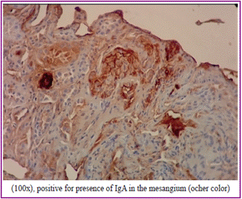
Four days later, he attended an appointment with an elevation of creatinine at 4.01 mg/dL and positive p-ANCAS, confirmed by ELISA (Myeloperoxidase, MPO 11.6) (Table 2). A renal biopsy was performed, documenting 6 glomeruli upon light microscopy, with mild endocapillary proliferation (polymorphonuclear), cellular extracapillary proliferation in more than half of the glomeruli, inflammatory cells (lymphocytes and plasma cells) in the interstitium, and mild tubular atrophy. No mesangial or vascular changes were evident (Figure 2). It was not possible to evaluate immunofluorescence due to lack of material, but immunohistochemistry was performed confirming IgA deposits in the mesangium (Figure 3). No electronic microscopy was available. The patient was considered to have AN-CA-associated RPGN, without extrarenal systemic manifestations of vasculitis.
Considering the histological findings and renal deterioration, despite having received a dose of cyclophosphamide, we decided to continue with cytostatic and steroids, and initiate plasmapheresis. Seven sessions of plasmapheresis were performed, showing a decrease in creatinine to 2.74 mg/dL and resulting in hospital discharge (Figure 1).
The patient changed his country of residence and therefore his treating doctor. Information was received from the patient about the continuity of cyclophosphamide and steroids. One month after hospital discharge, tests were performed to document stability of renal function, but persistence of hypoalbuminemia, proteinuria and hematuria (creatinine 2.39 mg/dL, BUN 36 mg/dL, albumin 2.9 gr/dL and urinalysis with 3+ of proteins and hematuria).
Literature review
IgAN is the most frequent primary glomerulonephritis, characterized by mesangial deposit of IgA. The rate of progression of the disease is varia ble; however, at 25 years of follow-up, 30-50% of patients progress to stage 5 chronic kidney disease (CKD5).2 Rapid progression has been reported in less than 10% of patients, with histological findings suggestive of vasculitis such as crescents, fibrinoid necrosis, and arteriolar lesion in some of them3.
The coexistence of antineutrophil cytoplasmic antibodies (ANCA) and IgAN is rare, being reported in 0.2-2 % of cases4. ANCA, mainly ANCA-myeloperoxidase (MPO), has been documented both in IgAN patients with histological findings of rapidly progressive glomerulonephritis, and in patients without these findings3-5. Screening for ANCA in patients with pauci-immune vasculitis is generally limited to the study of immunoglobulin G; however, ANCA of IgA class has been found in certain conditions, such as Henoch-Shonlein purpura, IgAN, among others6,7. The presence of IgA ANCA in crescentic IgAN (cIgAN) and pauci-immune crescentic glomerulonephritis (cGN) has also been reported8-10. Although the clinical behavior of patients with IgAN and ANCA has been evaluated, the pathogenic role thereof is still unclear, since it could be the hyperactivity of antibody producing cells or another concomitant disease3. It is even less clear if the presence of IgG or IgAANCA has any differential effect.
Several reports and case series of patients with IgAN and positive ANCA have been published11-16. Bantis et al. evaluated the behavior of 8 patients with crescentic IgAN and positive ANCA and compared them with 26 patients with more than 10 % of glomeruli with crescents, but negative ANCA. All patients with ANCA presented rapidly progressive glomerulonephritis, reaching a higher level of creatinine in the first three months than patients without ANCA (2.2-4.2 mg/dL vs. 1.9-2.5 mg/ dL). Histologically, the positivity for ANCA was related to a higher percentage of crescentic glomeruli (54% vs. 18 %)15.
Yang et al. carried out a retrospective study in which they evaluated the clinical and histological characteristics of 20 patients with IgAN and ANCA and compared them with 40 patients with IgAN-negative ANCA and 40 patients with systemic vasculitis associated with ANCA16. Patients with positive IgAN-ANCA, as well as those with ANCA vasculitis, were older (between 50-60 years old) and more frequently presented general symptoms and pulmonary compromise than the IgAN-negative ANCA group. No differences were found in the percentage of glomeruli with crescents, but there was a higher percentage of fibrinoid necrosis in IgAN-positive ANCA and ANCA vasculitis. Nine of 20 patients with IgAN-ANCA had cIgAN (crescents in >50% of glomeruli). The patients with cGN were compared in the three groups, finding similar age in cIgAN with and without ANCA, but older age compared with cGN-ANCA (54-59 vs. 40 years old, p=0.001). Systemic symptoms and lung com promise were more frequent in cIgAN-ANCA than in cIgAN-negative ANCA. No differences were found in the glomerular filtration rate (GFR) in the three groups. Dialysis was suspended in 75% of pa tients (3/4) with cIgAN-ANCA, compared to 9.1% of patients (1/11) with cIgAN-negative ANCA (p = 0.03), and less progression to CKD5 was evidenced at 6 months in cIgAN-ANCA. However, no differences were documented in this last outcome at the end of the follow-up (p = 0.11)16. This study concludes that patients with IgAN-ANCA present mixed characteristics of IgAN and ANCA vasculitis and have a more aggressive clinical and histological course than patients with IgAN-negative ANCA. We must carefully analyze the outcomes of dialysis suspension and progression to CKD5, since in this study ANCA negative patients received immunosuppressive treatment less frequently and had a higher proportion of sclerotic glomeruli in the biopsies, which may affect the interpretation of results.
Although the appearance of IgANc is rare, its aggressive course justifies knowing the factors that predict the renal prognosis. Jicheng Lv et al. are evaluating the long-term outcomes of a cohort of 113 patients with cIgAN (ANCA negative) and constructing a prediction model of renal recovery. The average serum creatinine, at the time of biopsy, was 4.3 +/- 3.4 mg/dL. The renal survival, at 1.3 and 5 years after the biopsy, was 57.4%, 45, 8% and 30.4%, respectively. The multivariate analysis documents that the initial serum creatinine is the only independent factor associated with CKD5 (HR 1.32, 95% CI, 1.1-1.57, p = 0.002), without finding an effect of the percentage of crescentic glomeruli. The risk of CKD5, one year after the biopsy, increases rapidly with creatinine of 2.7 mg/dL, and is 90% with creatinine of 6.8 mg/dL (sensitivity 98.5%, specificity 64.6%). With the latter, the possibility of renal recovery is lower, even with aggressive immunosuppressive treatment17.
There are no randomized studies that allow us to know which the ideal treatment for cIgAN is, but cases and series reported in the literature, where the treatment of other cGN and pauci-immune vasculitis is extrapolated, such as the use of steroids and cyclophosphamide, with variable results4,16-18. In fact, the 2011 KDIGO guidelines suggest not treating IgAN patients with corticosteroids combined with cyclophosphamide or azathioprine, unless they have cIgAN and rapid deterioration of renal function (recommendation 2D) 19. Plasmapheresis has been used in patients with cIgAN. The authors carrying out the aforementioned study on the prediction model of renal recovery17 establish plasmapheresis in their clinical practice in cIgAN patients with creatinine greater than 6.8 mg / dL (point of no return documented in their initial analysis), who require dialysis or with persistent deterioration of renal function despite receiving steroids or other immunosuppressants. They published the results of 12 patients with plasmapheresis, compared with a control group chosen by propensity analysis. During the follow-up (15 monthson average), 50 % of patients with plasmapheresis were free of dialysis compared to 0 % of the control group20. Additionally, a decrease in circulating IgA-IgG immunocom-plexes and complement activation products was documented20.
The association between IgAN and complement activation should also be mentioned, since mutations and polymorphisms of genes encoding factor H and factor H-related proteins have been found in this group of patients3. The deposit of C3 and C4, in kidney biopsies of IgAN, suggests the activation of the alternative pathway and lectins. From the cli nical point of view, the deposit of C4d in the mesangium and C3d in peritubular tumors have been associated with aggressive forms of the disease21,22. Complement blockers have already been evaluated in patients with lupus nephritis and ANCA vasculitis, and the use of eculizumab in a patient with IgA refractory to steroids, cyclophosphamide and plasmapheresis was reported23-25. Surely, until prospective and randomized studies are carried out to evaluate the effectiveness of this treatment in patients with cIgAN, steroids, other immunosuppressants and plasmapheresis will continue to be considered as some therapeutic options in this high-risk group.
Discussion
The case presented highlights the different clinical forms of IgAN, since the patient had CKD and, subsequently, nephrotic syndrome and acute kidney injury. While the biopsy was of little material, it was possible to demonstrate the presence of crescents, concordant with the clinical behavior of the patient. It was not possible to perform immunofluorescence, but the immunohistochemistry was positive for IgA in the mesangium, corroborating the previously diagnosed IgA. Based on the acute and severe deterioration of renal function, steroids and cyclophosphamide were initiated, as described in some case series referenced. Subsequently, due to the documentation of crescents, the positivity of ANCA-MPO and the elevation of serum creatinine, plasmapheresis was initiated, thus stopping the deterioration of renal function, but with persistence of nephrotic proteinuria. The short time of patient follow-up does not allow us to know the outcome.
Based on the review of the literature, it should be noted that, although the pathogenic role of ANCA in IgAN is unclear, the renal deterioration of this patient coincides with the appearance of crescents and ANCA not previously documented. No fibrinoid necrosis was found (one of the characteristic findings of ANCA vasculitis), nor did the patient report systemic symptoms or pulmonary compromise, as these patients do. Although the presence of fibrinoid necrosis is frequent in biopsies of patients with cIgAN-ANCA, not all cases reported in the literature present this finding16.
To date, there is not enough evidence to define the true meaning of ANCA in patients with IgAN. However, data from previously described studies suggest that patients with IgAN-ANCA have similar clinical characteristics to patients with ANCA vasculitis, have histological findings different from those in IgAN-negative ANCA, and frequently have cGN. The renal prognosis of patients with cIgAN-ANCA, compared to cIgAN-negative ANCA, is not clear, to the extent that the studies carried out so far do not allow to conclude exact data on this subject. cGN in IgA has been defined differently in the studies (different percentage of glomeruli with crescents) and the response to specific treatments compared with a control group has not been evaluated. In descriptive studies, patients with cIgAN-ANCA have been treated with immunosuppressive schemes used in patients with ANCA vasculitis, whose clinical response has been documented in some cases15.
Although there is not enough scientific evidence to predict the renal outcome in our patient, he is likely to present progression, considering his baseline renal function, persistent proteinuria, and the level of creatinine reached. This case shows an aggressive course of the disease, with a rare form such as cGN, and in which the finding of ANCA makes us wonder whether it is a coincidence or an additional disease.
Conclusion
IgA nephropathy is a disease that can eventually have an aggressive course, with rapidly progressive glomerulonephritis and coexistence of ANCA. The pathogenic role of ANCA in IgA nephropathy is unclear. However, the fact that patients with ANCA are clinically and histologically different from AN-CA negative patients, in the case series described in this text, suggests that they may be different entities.
It is important to evaluate the presence of ANCA in patients with IgAN of unusual course, accelerated deterioration and/or with crescents in the renal biopsy. If cIgAN and ANCA are documented, we suggest looking for extrarenal manifestations of vasculitis and initiating aggressive immunosuppressive treatment, as the case may be. It is expected that, with the conduct of studies in the future, we will be able to know which the most appropriate diagnostic approach and therapeutic intervention in this group of patients is.
Ethical disclosures
Protection of people and animals
The authors state that no human or animal experiments have been performed for this research.
Right to privacy and informed consent
The authors state that no patient data is included in this article.
Contribution of authors
Jorge Echeverri, Adalberto Pecha, and Carolina Larrarte: reviewed the patient's medical history, updated the information, reviewed the corresponding literature, and wrote the article.
Luis Javier Ossa, José de Jesús Arias, and Diana Gutiérrez: reviewed the histology plates of the renal biopsy, reviewed the case, and reported the pathology in the paper.
All the authors reviewed the writing at the end and approved the result.


 text in


